. 2012 Apr 1;50(3):725-33.
doi: 10.1016/j.ijbiomac.2011.12.012. Epub 2011 Dec 23.

Toxoplasma gondii Sis1-like J-domain protein is a cytosolic chaperone associated to HSP90/HSP70 complex

Affiliations

Toxoplasma gondii Sis1-like J-domain protein is a cytosolic chaperone associated to HSP90/HSP70 complex

Maria J Figueras et al. Int J Biol Macromol. .

Abstract

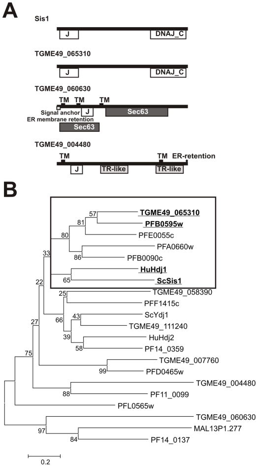
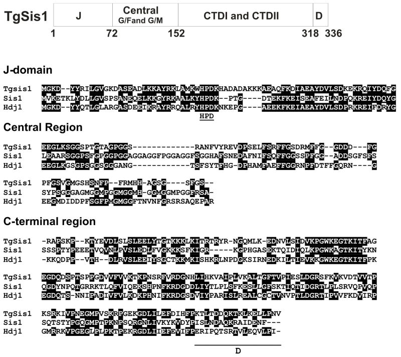
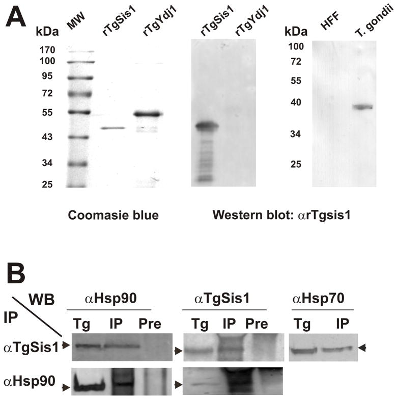
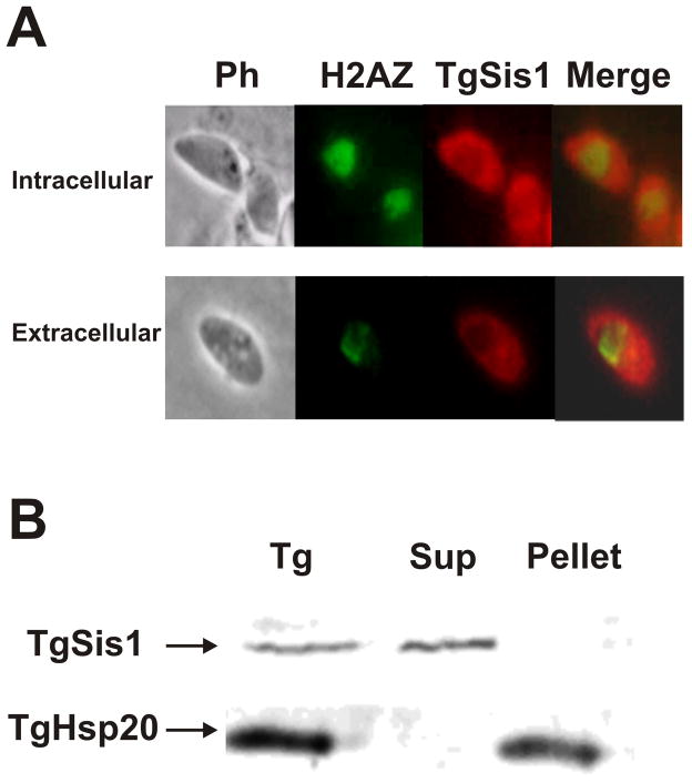
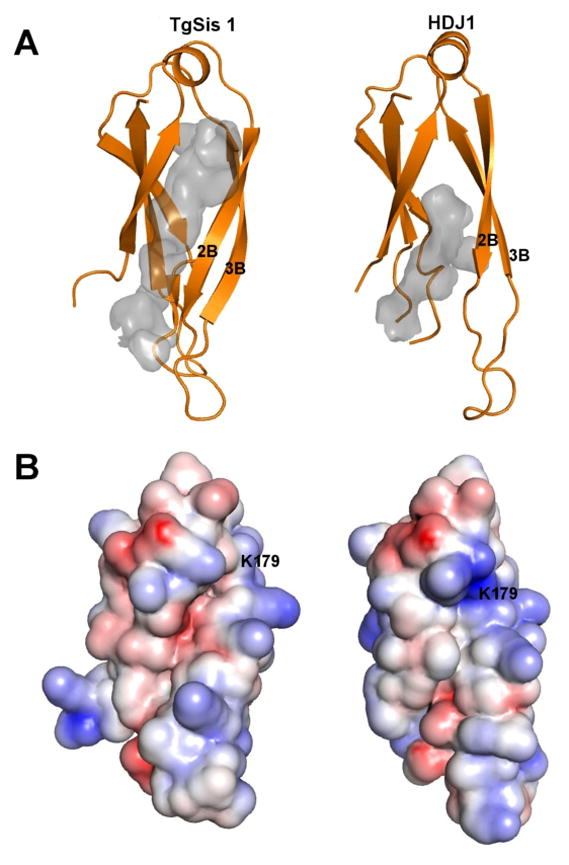
Toxoplasma gondii is an obligate intracellular protozoan parasite in which 36 predicted Hsp40 family members were identified by searching the T. gondii genome. The predicted protein sequence from the gene ID TGME49_065310 showed an amino acid sequence and domain structure similar to Saccharomyces cerevisiae Sis1. TgSis1 did not show differences in its expression profile during alkaline stress by microarray analysis. Furthermore, TgSis1 showed to be a cytosolic Hsp40 which co-immunoprecipitated with T. gondii Hsp70 and Hsp90. Structural modeling of the TgSis1 peptide binding fragment revealed structural and electrostatic properties different from the experimental model of human Sis1-like protein (Hdj1). Based on these differences; we propose that TgSis1 may be a potentially attractive drug target for developing a novel anti-T. gondii therapy.


Figures

Fig. 1
Fig. 1
Domain organization and phylogenetic analysis of T. gondii Type I and II J-domain proteins. (A) Domain and motif organization of yeast Sis1 and T. gondii putative type II Hsp40s. Domains and motifs were identified by NCBI (http://www.ncbi.nlm.nih.gov/Structure/cdd/wrpsb.cgi) and Motif Scan (http://myhits.isb-sib.ch/cgi-bin/motif_scan). (B) Neighbor joining (NJ) tree of Type I and II J-domain protein amino acid sequences of T. gondii, P. falciparum, S. cerevisiae Sis1, and human Hdj1. T. gondii sequences were included on the basis of gene ID release 4 (see Table 1). P. falciparum gene IDs begin with PF except Malp13P1.277. Frame indicates cluster of sequences related to Sis1/Hdj1. Type I and II sequences were analyzed because Sis1-like Hsp40 share the CTD domain present in type I Hsp40s.
Fig. 2
Fig. 2
Sequence analysis of T. gondii Sis1 like protein (TgSis1). Upper panel: Domains of TGME49_065310 (putative TgSis1). J: J-domain; Central: central region that includes G/F- and G/M-rich regions; C-terminal I/II: C-terminal domain (CTD) containing Domain I (peptide binding fragment) and Domain II; D: dimerization motif. Middle and bottom panels: Sequence alignment generated by clustal W (Bioedit program). Letters highlighted in black indicate identical residues. Gaps are indicated by dashes and were introduced to improve the alignment. Sis1, S. cereviseae Sis1 (AN: CAA41366), HuHDJ1, Human HDJ1 (AN: P25685). The sequence was split in J-domain, Central region and C-terminal region (containing CTDI, CTDII and dimerization motif [underlined]) according Lee et al., [22] full length alignment.
Fig. 3
Fig. 3
Detection of native TgSis1-like protein and co-immunoprecipitation analysis. (A) Left panel, purified recombinant rTgSis1 and putative rTgYdj1 were electrophoresed in SDS-PAGE and stained with Coomassie Brilliant Blue. Middle and right panels, Western blot with rabbit α-TgSis1. T. gondii: parasite RH strain lysate. HFF: Protein extract from uninfected HFF cells. Pre-immune serum samples did not show reactivity (data not shown). Migration of molecular weight markers (PageRulerPrestainded Protein Ladder, Fermentas International Inc.) is indicated in kilo Daltons (kDa). (B) Co-immunoprecipitation analysis. T. gondii RH strain lysates were used for immunoprecipitation (IP) using specific α-TgSis1 and T. gondii Hsp90 (α–Hsp90) antisera as indicated on the left side. IPs were analyzed by Western blotting with αTgSis1, α-Hsp90, and a commercial anti-human Hsp70 antibody (αHsp70). T. gondii RH strain lysate (Tg) was included in the Western blot analysis to identify the corresponding band. IP with pre-immune sera (Pre) was also analyzed with αTgSis1 and αHsp90 to test the specificity of the IP.
Fig. 4
Fig. 4
Subcellular localization of TgSis1. (A) Indirect immunofluorescence assays (IFA) and subcellular localization of TgSis1-like protein in tachyzoites (Tz). IFA was performed on fixed intracellular parasites with rabbit αTgSis1 and murine anti-H2AZNt (H2AZ serves as a marker of the parasite’s nucleus). Merged, merge of TgSis1 and H2AZ images. (B) Tachyzoite lysate (Tg) was obtained by freeze/thaw in MOPS buffer, centrifuged to obtain the pellet and the supernatant (Sup). Samples were analyzed by Western blot with anti-T. gondii Hsp20 antibody and αTgSis1.
Fig. 5
Fig. 5
Structural comparison of peptide binding fragment (Domain I) of TgSis1 and of HjD1. (A) Both structures were drawn with PyMol using cartoon representation. It is clearly seen that the largest differences reside at the secondary structure and position of strand 2 and 3. The peptide binding sites are shown as gray cavities. The actual shape of the cavities depends on several user-adjustable parameters, but the peptide binding site of TgHSP40 is consistently deeper, a bit narrower and longer than the peptide binding site of Hdj1. (B) Electrostatic potential of Domain I of the peptide binding fragment of Hsp40 in PyMol plotted on the solvent-accessible surface from −5 kT/e (basic residue, Red) to +5 kT/e (acid residue, Blue).

Similar articles

Cited by

References

    1. Israelski DM, Chmiel JS, Poggensee L, Phair JP, Remington JS. J Acquir Immune Defic Syndr. 1993;6:414–418. - PubMed
    1. Luft BJ, Remington JS. Clin Infect Dis. 1992;15:211–222. - PubMed
    1. Black MW, Boothroyd JC. Microbiol Mol Biol Rev. 2000;64:607–623. - PMC - PubMed
    1. Tenter AM, Heckeroth AR, Weiss LM. Int J Parasitol. 2000;30:1217–1258. - PMC - PubMed
    1. Vonlaufen N, Kanzok SM, Wek RC, Sullivan WJ., Jr Cell Microbiol. 2008;10:2387–2399. - PubMed

Publication types

MeSH terms

LinkOut - more resources