

Microglial activation and TDP-43 pathology correlate with executive dysfunction in amyotrophic lateral sclerosis
- PMID: 22210083
- PMCID: PMC3595560
- DOI: 10.1007/s00401-011-0932-x
Microglial activation and TDP-43 pathology correlate with executive dysfunction in amyotrophic lateral sclerosis
Abstract
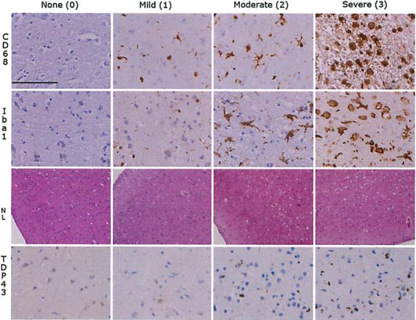
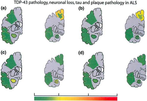
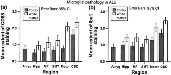
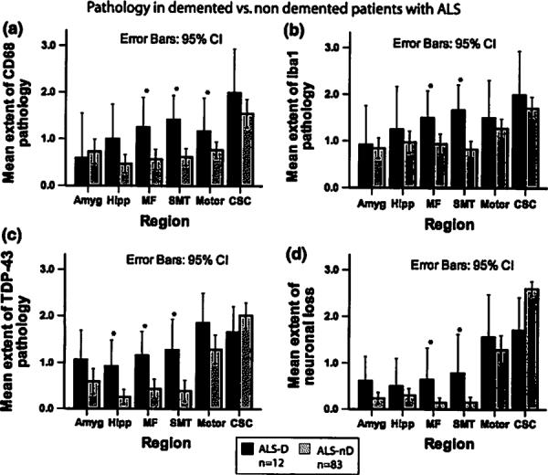
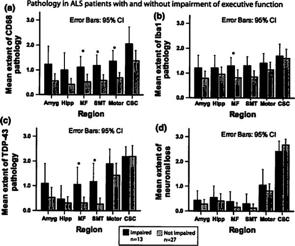
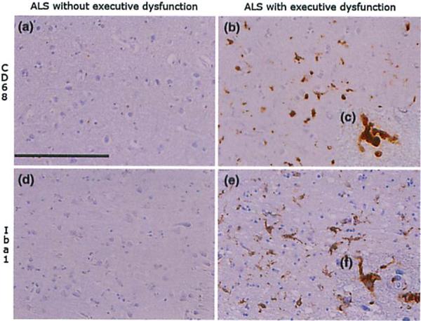
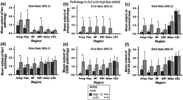
While cognitive deficits are increasingly recognized as common symptoms in amyotrophic lateral sclerosis (ALS), the underlying histopathologic basis for this is not known, nor has the relevance of neuroinflammatory mechanisms and microglial activation to cognitive impairment (CI) in ALS been systematically analyzed. Staining for neurodegenerative disease pathology, TDP-43, and microglial activation markers (CD68, Iba1) was performed in 102 autopsy cases of ALS, and neuropathology data were related to clinical and neuropsychological measures. ALS with dementia (ALS-D) and ALS with impaired executive function (ALS-Ex) patients showed significant microglial activation in middle frontal and superior or middle temporal (SMT) gyrus regions, as well as significant neuronal loss and TDP-43 pathology in these regions. Microglial activation and TDP-43 pathology in middle frontal and superior or middle temporal regions were highly correlated with measures of executive impairment, but not with the MMSE. In contrast, only one ALS-D patient showed moderate Alzheimer's disease (AD) pathology. Tau and Aβ pathology increased with age. A lower MMSE score correlated with tau pathology in hippocampus and SMT gyrus, and with Aβ pathology in limbic and most cortical regions. Tau and Aβ pathology did not correlate with executive measures. We conclude that microglial activation and TDP-43 pathology in frontotemporal areas are determinants of FTLD spectrum dementia in ALS and correlate with neuropsychological measures of executive dysfunction. In contrast, AD pathology in ALS is primarily related to increasing age and associated with a poorer performance on the MMSE.
Figures
References
-
- Abrahams S, Goldstein LH, Suckling J, Ng V, Simmons A, Chitnis X, Atkins L, Williams SC, Leigh PN. Frontotemporal white matter changes in amyotrophic lateral sclerosis. J Neural. 2005;252:321–331. - PubMed
-
- Alexianu ME, Kozovska M, Appel SH. Immune reactivity in a mouse model of familial ALS correlates with disease progression. Neurology. 2001;57:1282–1289. - PubMed
-
- Arai T, Hasegawa M, Nonoka T, Kametani F, Yamashita M, Hosokawa M, Niizato K, Tsuchiya K, Kobayashi Z, Ikeda K, Yoshida M, Onaya M, Fujishiro H, Akiyama H. Phosphorated and cleaved TDP-43 in ALS, FTLD and other neurodegenerative disorders and in cellular models of TDP-43 proteinopathy. Neuropathology. 2010;30:170–181. - PubMed
Publication types
MeSH terms
Substances
Grants and funding
LinkOut - more resources
Full Text Sources
Medical
Miscellaneous
