. 2012 Jan 24;109(4):1127-32.
doi: 10.1073/pnas.1111053108. Epub 2011 Dec 30.

CD146, an epithelial-mesenchymal transition inducer, is associated with triple-negative breast cancer

Affiliations

CD146, an epithelial-mesenchymal transition inducer, is associated with triple-negative breast cancer

Qiqun Zeng et al. Proc Natl Acad Sci U S A. .

Abstract

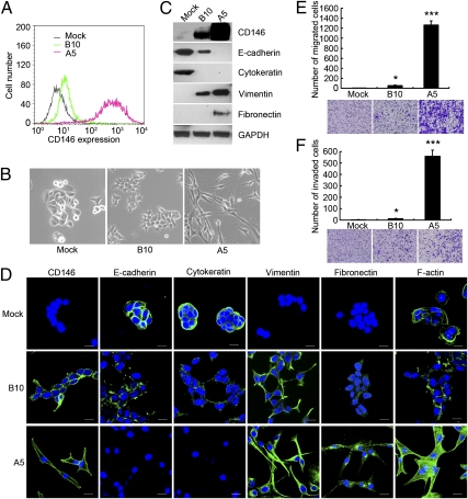
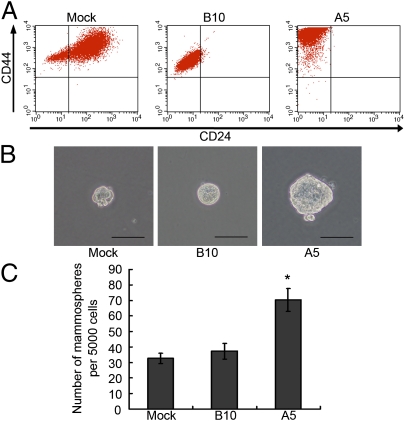
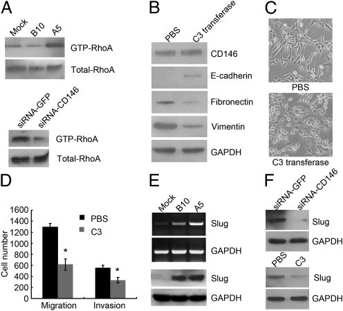
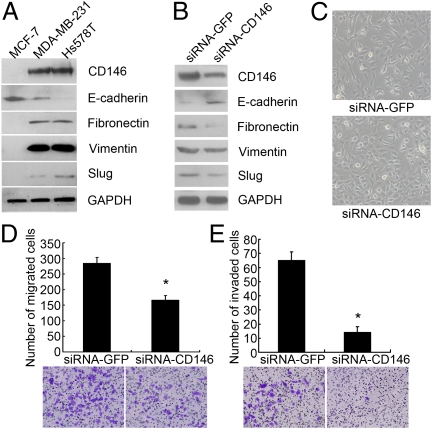
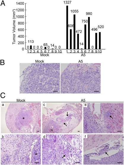
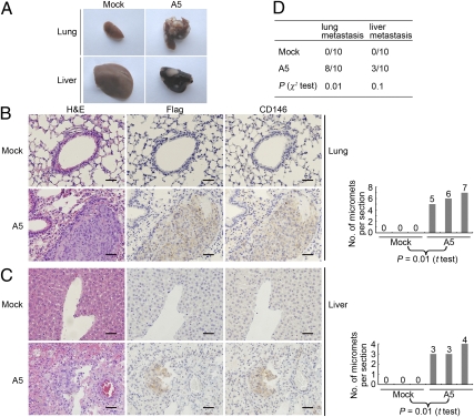
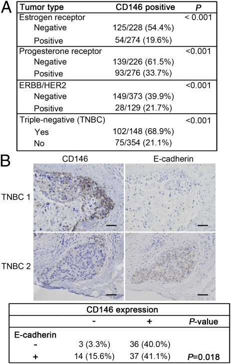
The epithelial-mesenchymal transition (EMT) plays an important role in breast cancer metastasis, especially in the most aggressive and lethal subtype, "triple-negative breast cancer" (TNBC). Here, we report that CD146 is a unique activator of EMTs and significantly correlates with TNBC. In epithelial breast cancer cells, overexpression of CD146 down-regulated epithelial markers and up-regulated mesenchymal markers, significantly promoted cell migration and invasion, and induced cancer stem cell-like properties. We further found that RhoA pathways positively regulated CD146-induced EMTs via the key EMT transcriptional factor Slug. An orthotopic breast tumor model demonstrated that CD146-overexpressing breast tumors showed a poorly differentiated phenotype and displayed increased tumor invasion and metastasis. We confirmed these findings by conducting an immunohistochemical analysis of 505 human primary breast tumor tissues and found that CD146 expression was significantly associated with high tumor stage, poor prognosis, and TNBC. CD146 was expressed at abnormally high levels (68.9%), and was strongly associated with E-cadherin down-regulation in TNBC samples. Taken together, these findings provide unique evidence that CD146 promotes breast cancer progression by induction of EMTs via the activation of RhoA and up-regulation of Slug. Thus, CD146 could be a therapeutic target for breast cancer, especially for TNBC.


Conflict of interest statement

The authors declare no conflict of interest.

Figures

Fig. 1.
Fig. 1.
CD146 induces EMTs in human breast cancer cells. (A) FACS analysis of CD146 expression in vector control MCF-7-Mock (Mock) and CD146 clones MCF-7-B10 (B10) and MCF-7-A5 (A5). (B) Morphology of Mock, B10, and A5 cells. Magnification, 200×. (C and D) Expression of CD146 and EMT markers analyzed by immunoblotting and immunofluorescence. Nuclei are shown with DAPI staining. (Scale bars, 20 μm.) (E and F) Migration and invasion assays of Mock, B10, and A5 cells. Data were collected from three wells, *P < 0.05, ***P < 0.001, compared with Mock cells. Representative images of migrated or invaded cells are also shown.
Fig. 2.
Fig. 2.
CD146-induced EMTs generate breast CSC-like cells. (A) FACS analysis of cell-surface markers CD44 and CD24 in Mock, B10, and A5 cells. (B and C) Morphology and quantification of mammospheres formed by Mock, B10, and A5 cells. (Scale bars, 100 μm.) *P < 0.05, compared with Mock cells.
Fig. 3.
Fig. 3.
RhoA activation and Slug up-regulation mediate CD146-induced EMTs. (A) GTP-RhoA affinity pull-down assay to determine the level of GTP-RhoA in Mock, B10, and A5 (Upper), and the level of GTP-RhoA after CD146 silencing in A5 cells (Lower). (B) Immunoblotting of CD146 and EMT markers in A5 cells after PBS or C3 transferase treatment (1 μg/mL, 24 h). (C) Morphology of A5 cells after PBS or C3 transferase treatment. Magnification, 100×. (D) Migration and invasion assays in A5 cells after PBS or C3 transferase treatment. *P < 0.05, compared with the control. (E) Slug expression in Mock, B10, and A5 cells analyzed by RT-PCR (Upper) and immunoblotting (Lower). (F) Slug expression in A5 cells after CD146 silencing (Upper) or C3 transferase treatment (Lower) analyzed by immunoblotting.
Fig. 4.
Fig. 4.
Down-regulation of CD146 expression reverses the mesenchymal phenotype of MDA-MB-231 cells. (A) Immunoblotting of CD146 and EMT markers in MCF-7, Hs578T, and MDA-MB-231 cells. (B) Immunoblotting of CD146, E-cadherin, fibronectin, vimentin, and Slug after CD146 silencing in MDA-MB-231 cells. (C) Morphology of MDA-MB-231 cells after CD146 silencing. Magnification, 100×. (D and E) Migration and invasion assays of MDA-MB-231 cells after CD146 silencing. Data were collected from three wells, *P < 0.05, compared with the control. Representative images of migrated or invaded cells are also shown.
Fig. 5.
Fig. 5.
CD146 promotes tumor invasion in vivo. (A) Individual volume of Mock and A5 tumors at week 10 after orthotopic injection. (B) Mock and A5 tumor sections stained with H&E. (Scale bars, 100 μm.) (C) H&E staining in Mock and A5 tumor sections and adjacent tissues. (a and b) Mock tumors without stromal invasion. (c and d, arrows) Areas of stromal invasion of A5 tumors. (e and f, arrows) Areas of muscular and vascular invasion of A5 tumors. Asterisks, Tumor mass. N, necrosis. (Scale bars, 100 μm.)
Fig. 6.
Fig. 6.
CD146 promotes breast tumor metastasis in vivo. (A) Morphology of lung and liver metastases formed by Mock and A5 cells at week 10 after orthotopic injection. (B and C) H&E-, Flag-, and CD146-stained section of lungs and livers isolated from mice carrying Mock and A5 tumors. Arrows indicate representative micrometastases (Left). Numbers of micrometastases (micromets) per section in lung and livers in individual mice carrying Mock and A5 tumors (Right). Data were collected from three mice per group. t Tests were used to determine differences. P < 0.05 was considered to be significant. (Scale bars, 100 μm.) (D) Incidence of lung and liver metastasis in mice carrying Mock and A5 tumors, as analyzed by morphology observation and immunohistochemistry. The Pearson χ2 test was used to determine correlations. P < 0.05 were considered significant.
Fig. 7.
Fig. 7.
CD146 is associated with triple-negative breast cancer. (A) Associations of CD146 expression with ER-, PR-, HER2-statuses, and with the triple-negative phenotype in 502 human breast tumor tissues, for which ER, PR, and HER2 status were available. (B) Representative immunohistochemical staining of CD146 and E-cadherin in 2 TNBC samples (Upper), and statistical correlations between CD146 expression and E-cadherin in TNBC (Lower). Only membrane expression of E-cadherin was considered to be positive. The Pearson χ2 test was used to determine correlations. P < 0.05 were considered significant. (Scale bars, 100 μm.)

References

    1. Jemal A, et al. Global cancer statistics. CA Cancer J Clin. 2011;61:69–90. - PubMed
    1. Thiery JP, Acloque H, Huang RY, Nieto MA. Epithelial-mesenchymal transitions in development and disease. Cell. 2009;139:871–890. - PubMed
    1. Mani SA, et al. The epithelial-mesenchymal transition generates cells with properties of stem cells. Cell. 2008;133:704–715. - PMC - PubMed
    1. Kang Y, Massagué J. Epithelial-mesenchymal transitions: Twist in development and metastasis. Cell. 2004;118:277–279. - PubMed
    1. Yilmaz M, Christofori G. EMT, the cytoskeleton, and cancer cell invasion. Cancer Metastasis Rev. 2009;28:15–33. - PubMed

Publication types

MeSH terms