. 2012 Sep;2(3):113-20.
doi: 10.1007/s12348-011-0054-6. Epub 2012 Jan 1.

Characterization of macular lesions in punctate inner choroidopathy with spectral domain optical coherence tomography

Affiliations

Characterization of macular lesions in punctate inner choroidopathy with spectral domain optical coherence tomography

Roomasa Channa et al. J Ophthalmic Inflamm Infect. 2012 Sep.

Abstract

Purpose: Punctate inner choroidopathy (PIC) is an ocular inflammatory disease. Spectral domain optical coherence tomography (SD-OCT) allows detailed visualization of retinal and choroidal structures. We aimed to describe the retinal changes on SD-OCT associated with PIC lesions localized in the macula.

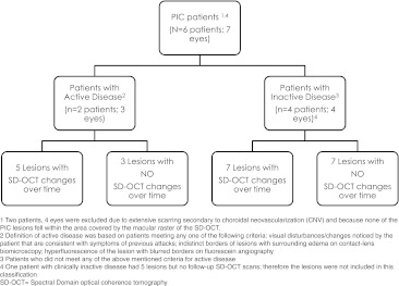
Methods: Retrospective case series: PIC lesions not associated with choroidal neovascularization (CNV) and captured by macular SD-OCT scans were identified and characterized.

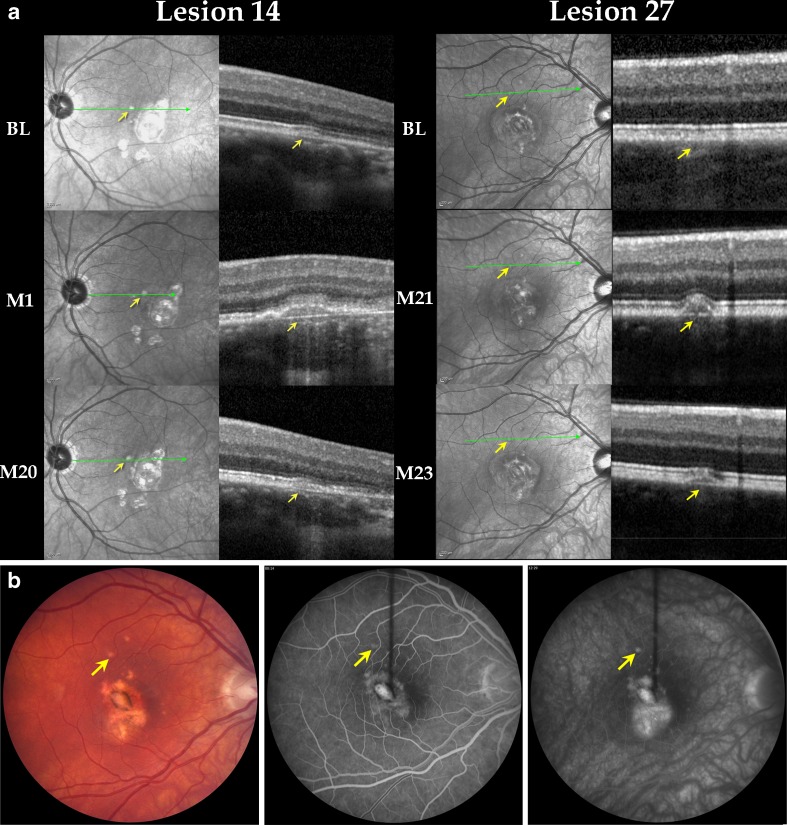
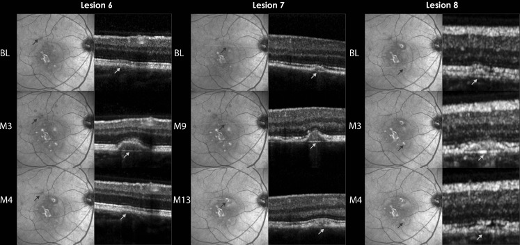
Results: Twenty-seven PIC lesions from seven patients (eight eyes) were identified and classified into four categories according to disease activity and temporal changes. Among clinically inactive patients, two main patterns were noted on OCT: (1) retinal pigment epithelium (RPE) elevation with sub-RPE hyper-reflective signals and (2) localized disruption of outer retinal layers with choroid and Bruch's membrane (BM) generally spared. Clinically active patients demonstrated lesions with intact BM with RPE elevation that fluctuated with disease activity and sub-RPE hyper-reflective signals. Photoreceptor-associated bands on SD-OCT (PRs) were not visible during active disease, but returned to normal visibility when lesions were clinically stable. Seven lesions in patients without clinically detected activity demonstrated alteration of RPE elevation.

Conclusion: SD-OCT can provide detailed structural characteristics of PIC lesions. RPE elevation is noted in many lesions while BM and choroid are spared. Photoreceptor-associated bands on SD-OCT appear compressed during clinically active stages and are visible during stabilization. OCT may provide information on activity not detected clinically.


Figures

Fig. 1
Fig. 1
Categorization of PIC lesions in patients with active and inactive disease
Fig. 2
Fig. 2
a Lesions in clinically active patients with temporal changes on spectral domain optical coherence tomography is a composite of IR and OCT images. The arrows point towards the lesion. The first row shows IR and OCT images at BL, the middle row shows IR and OCT images at the time of clinical activity, and the bottom row shows IR and OCT images of the same lesion at the last follow-up visit after resolution of clinical activity. Left (lesion 14): lesion (in the left eye) was initially seen as a small RPE elevation. Patient was clinically active at M1; during active disease, PRs were not visible. Resolution of RPE elevation and reappearance PRs were seen at M20. Right (lesion 27): lesion (in the right eye) was first seen at the time of clinical activity (M21) as a small RPE elevation, and PRs were partially visible. Two months later, the RPE elevation had decreased. BL first visit for which SD-OCT was available, M month, PR photoreceptor-associated bands on SD-OCT. b Color fundus photograph (left), early FA (center), and late FA (right) from Patient 7. The lesion marked by the arrows corresponds to lesion 27 as it appeared on color fundus and FA at M21 during clinical activity. The late FA shows mild leakage with blurring of the borders of the lesion. FA fluorescein angiogram, M month, IR infrared
Fig. 3
Fig. 3
Lesions in clinically stable patient with temporal changes on spectral domain optical coherence tomography is a composite of IR and OCT images showing three lesions from the right eye of Patient 2. All three lesions (6, 7, and 8) showed retinal pigment epithelium (RPE) elevation with disappearance of PRs, followed by resolution of the RPE elevation and reappearance of PRs. BL time point when lesion was first observed on SD-OCT, M month, PRs photoreceptor-associated bands on SD-OCT, IR infrared
Fig. 4
Fig. 4
Clinically stable lesions that have remained unchanged on spectral domain optical coherence tomography scans during multiple visits. This figure shows magnified SD-OCT scans. The arrows point to the lesions. Top left (lesion 19): focal retinal pigment epithelium (RPE) disruption remained unchanged over a period of 16 months. Top right (lesion 2): focal irregularity of RPE and photoreceptor IS/OS junctions with choroidal thinning, unchanged over a period of 8 months. Bottom left (lesion 20): RPE elevation with sub-RPE signal, unchanged over a period of 16 months. Bottom right (lesion 15): focal disruption of photoreceptor IS/OS junctions with thinning of the ONL, unchanged over a period of 6 months. Repeated OCT scans at subsequent visits did not change in appearance compared to the images illustrated in these figures, and therefore, are not shown. SD-OCT spectral domain optical coherence tomography, RPE retinal pigment epithelium, IS/OS inner segment/outer segment, ONL outer nuclear layer, mos months

References

    1. Watzke RC, et al. Punctate inner choroidopathy. Am J Ophthalmol. 1984;98(5):572–584. - PubMed
    1. Akman A, Kadayifcilar S, Aydin P. Indocyanine green angiographic findings in a case of punctate inner choroidopathy. Eur J Ophthalmol. 1998;8(3):191–194. - PubMed
    1. Tiffin P, et al. Indocyanine green angiography in a case of punctate inner choroidopathy. Br J Ophthalmol. 1996;80(1):90. doi: 10.1136/bjo.80.1.90. - DOI - PMC - PubMed
    1. Turkcuoglu P, et al. Mycophenolate mofetil and fundus autofluorescence in the management of recurrent punctate inner choroidopathy. Ocul Immunol Inflamm. 2011;19(4):286–292. doi: 10.3109/09273948.2011.580072. - DOI - PubMed
    1. Hassenstein A, Meyer CH. Clinical use and research applications of Heidelberg retinal angiography and spectral-domain optical coherence tomography—a review. Clin Exp Ophthalmol. 2009;37(1):130–143. doi: 10.1111/j.1442-9071.2009.02017.x. - DOI - PubMed

LinkOut - more resources