. 2012 Feb;61(2):513-23.
doi: 10.2337/db11-0313. Epub 2011 Dec 30.

Histone deacetylase 6 (HDAC6) is an essential modifier of glucocorticoid-induced hepatic gluconeogenesis

Affiliations

Histone deacetylase 6 (HDAC6) is an essential modifier of glucocorticoid-induced hepatic gluconeogenesis

Robin Winkler et al. Diabetes. 2012 Feb.

Abstract

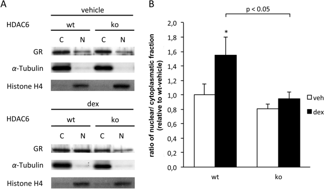
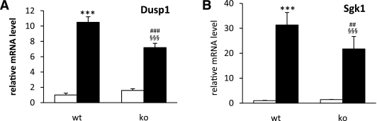
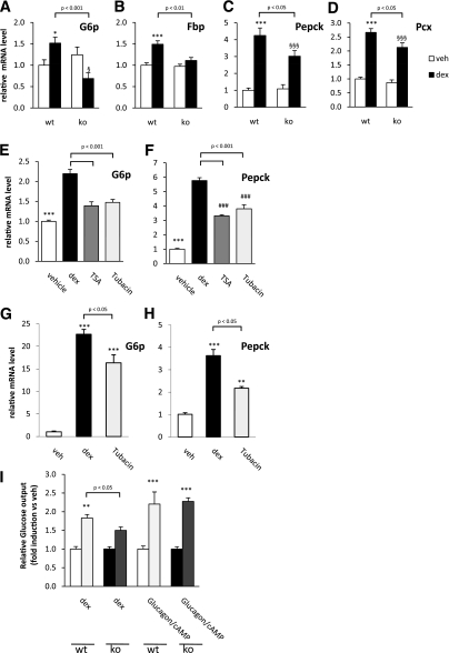
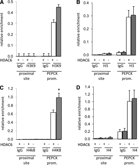
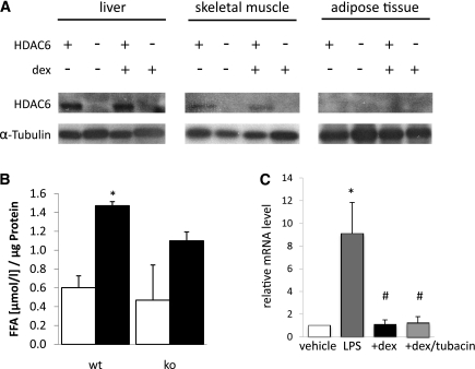
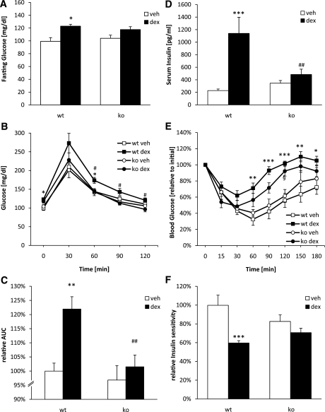
In the current study, we investigated the importance of histone deacetylase (HDAC)6 for glucocorticoid receptor-mediated effects on glucose metabolism and its potential as a therapeutic target for the prevention of glucocorticoid-induced diabetes. Dexamethasone-induced hepatic glucose output and glucocorticoid receptor translocation were analyzed in wild-type (wt) and HDAC6-deficient (HDAC6KO) mice. The effect of the specific HDAC6 inhibitor tubacin was analyzed in vitro. wt and HDAC6KO mice were subjected to 3 weeks' dexamethasone treatment before analysis of glucose and insulin tolerance. HDAC6KO mice showed impaired dexamethasone-induced hepatic glucocorticoid receptor translocation. Accordingly, dexamethasone-induced expression of a large number of hepatic genes was significantly attenuated in mice lacking HDAC6 and by tubacin in vitro. Glucose output of primary hepatocytes from HDAC6KO mice was diminished. A significant improvement of dexamethasone-induced whole-body glucose intolerance as well as insulin resistance in HDAC6KO mice compared with wt littermates was observed. This study demonstrates that HDAC6 is an essential regulator of hepatic glucocorticoid-stimulated gluconeogenesis and impairment of whole-body glucose metabolism through modification of glucocorticoid receptor nuclear translocation. Selective pharmacological inhibition of HDAC6 may provide a future therapeutic option against the prodiabetogenic actions of glucocorticoids.


Figures

FIG. 1.
FIG. 1.
Impaired glucocorticoid-induced glucocorticoid receptor (GR) translocation attributed to lack of HDAC6. A: Nuclear (N) and cytoplasmic (C) protein fractions of livers from wt and HDAC6KO (ko) mice were Western blotted for glucocorticoid receptor. Representative immunoblots from three sets of animals are shown. B: Densitometric analysis from HDAC6 Western immunoblots as the ratio of nuclear to cytoplasmatic fraction. (See A.) *P < 0.05 vs. wt plus vehicle (veh). dex, dexamethasone.
FIG. 2.
FIG. 2.
Attenuation of dexamethasone-induced hepatic mRNA regulation in HDAC6KO (ko) mice (A) and real-time quantitative PCR analysis of Dusp1 and Sgk1 showing attenuated dexamethasone-induced gene regulation in HDAC6KO mice (B). Data represent means ± SEM (n = 7–8). ***P < 0.001 vs. wt plus vehicle, ###P < 0.001 vs. HDAC6KO plus vehicle, §§§P < 0.001 vs. wt plus vehicle. ■, dexamethasone; □, vehicle.
FIG. 3.
FIG. 3.
Regulation of gluconeogenic genes by dexamethasone (dex) in the liver is attenuated in HDAC6KO (ko) mice. AD: Real-time quantitative PCR analysis of G6p, Fbp, Pepck, and Pcx of animal liver mRNA (n = 7–8 mice). E and F: H4IIE cells were stimulated with dexamethasone (500 nmol/L) for 4 h after 30-min pretreatment with vehicle (veh) and HDAC inhibitors (5 μmol/L tubacin and 1 μmol/L trichostatin A [TSA]). Real-time quantitative PCR analysis was conducted for the respective genes. G and H: Primary murine hepatocytes were stimulated with dexamethasone (500 nmol/L) after pretreatment with vehicle or tubacin (5 μmol/L). Real-time quantitative PCR analysis was conducted for the respective genes. Data represent means ± SEM of three independently conducted experiments. I: wt and HDAC6KO primary hepatocytes were treated with dexamethasone (10 nmol/L) and glucagon (100 nmol/L)/cAMP (1 μmol/L). Glucose content of the supernatant was quantified and normalized to total protein amount. Glucose output was conducted three times in triplicates. Data represent means ± SEM. AD: *P < 0.05, ***P < 0.001 vs. wt plus vehicle; §P < 0.05, §§§P < 0.001 vs. HDAC6KO plus vehicle. E\x{2013}F: ***P < 0.001, ###P < 0.001 vs. vehicle. G\x{2013}I: ***P < 0.001, **P < 0.01 vs. respective vehicle.
FIG. 4.
FIG. 4.
HDAC6 regulates hepatic histone acetylation. Chromatin immunoprecipitation (ChIP) analysis of the PEPCK promoter (prom.) shows histone H3 and H4 acetylation in livers from wt (+) and HDAC6KO (−) mice. Details can be found in research design and methods. The amount of acetylated H3K9 (A) and H4K8 (C) (proximal site: IgG, H4K8. PEPCK promoter [prom.]: IgG, no detectable signal after adequate amplification) and total H3 (B) and H4 (D) associated with the PEPCK promoter was analyzed. Results represent data from three independent experiments. *P < 0.05 vs. wt.
FIG. 5.
FIG. 5.
Regulation of glucocorticoid-mediated actions by HDAC6 in adipose tissue and monocytes. A: Western immunoblotting of HDAC6 in liver, skeletal muscle, and gonadal adipose tissue from wt (+) and HDAC6KO (−) mice injected with vehicle (−) or dexamethasone (+) according to the protocol described in research design and methods. B: Ex vivo lipolysis (free fatty acid [FFA] release) from gonadal fat pads injected with vehicle (□) or dexamethasone (■) (100 nmol/L). n = 5 wt and HDAC6KO mice were studied. *P < 0.05 vs. wt plus vehicle. C: IL-6 mRNA expression in human THP-1 cells after stimulation with vehicle or 10 ng/mL LPS for 13 h followed by treatment with dexamethasone (50 nmol/L) with or without tubacin (20 μmol/L). *P < 0.05 vs. vehicle, #P < 0.05 vs. LPS alone.
FIG. 6.
FIG. 6.
Dexamethasone (dex)-induced impaired glucose tolerance and insulin resistance are attenuated in HDAC6KO (ko) mice. A: Blood glucose values determined in wt and HDAC6KO mice after an overnight fast. B: GTT. After 3 weeks of treatment, mice were given an injection of 1 mg glucose/kg body wt i.p. after overnight fasting. C: Corresponding AUC. D: Serum insulin in the fed state. E: ITT. After 4 weeks of treatment, fasted mice were given an injection of 0.5 units of insulin/kg body wt i.p. Results represent blood glucose concentration as a percentage of the initial glucose value. F: Corresponding AUC. Data represent means ± SEM (n = 5–9). *P < 0.05, **P < 0.01, ***P < 0.001 vs. wt plus vehicle (veh), #P < 0.05 vs. HDAC6KO plus dexamethasone, ##P < 0.01 vs. wt plus dexamethasone, §P < 0.05 vs. HDAC6KO plus vehicle.

Comment in

Similar articles

Cited by

References

    1. Trikudanathan S, McMahon GT. Optimum management of glucocorticoid-treated patients. Nat Clin Pract Endocrinol Metab 2008;4:262–271 - PubMed
    1. Andrews RC, Walker BR. Glucocorticoids and insulin resistance: old hormones, new targets. Clin Sci (Lond) 1999;96:513–523 - PubMed
    1. Arner P, Gunnarsson R, Blomdahl S, Groth CG. Some characteristics of steroid diabetes: a study in renal-transplant recipients receiving high-dose corticosteroid therapy. Diabetes Care 1983;6:23–25 - PubMed
    1. Bernal-Mizrachi C, Weng S, Feng C, et al. Dexamethasone induction of hypertension and diabetes is PPAR-alpha dependent in LDL receptor-null mice. Nat Med 2003;9:1069–1075 - PubMed
    1. Delaunay F, Khan A, Cintra A, et al. Pancreatic beta cells are important targets for the diabetogenic effects of glucocorticoids. J Clin Invest 1997;100:2094–2098 - PMC - PubMed

Publication types

MeSH terms