. 2012 Mar;194(5):1113-26.
doi: 10.1128/JB.06549-11. Epub 2011 Dec 30.

Altered regulation of Escherichia coli biotin biosynthesis in BirA superrepressor mutant strains

Affiliations

Altered regulation of Escherichia coli biotin biosynthesis in BirA superrepressor mutant strains

Vandana Chakravartty et al. J Bacteriol. 2012 Mar.

Abstract

Transcription of the Escherichia coli biotin (bio) operon is directly regulated by the biotin protein ligase BirA, the enzyme that covalently attaches biotin to its cognate acceptor proteins. Binding of BirA to the bio operator requires dimerization of the protein, which is triggered by BirA-catalyzed synthesis of biotinoyl-adenylate (biotinoyl-5'-AMP), the obligatory intermediate of the ligation reaction. Although several aspects of this regulatory system are well understood, no BirA superrepressor mutant strains had been isolated. Such superrepressor BirA proteins would repress the biotin operon transcription in vivo at biotin concentrations well below those needed for repression by wild-type BirA. We isolated mutant strains having this phenotype by a combined selection-screening approach and resolved multiple mutations to give several birA superrepressor alleles, each having a single mutation, all of which showed repression dominant over that of the wild-type allele. All of these mutant strains repressed bio operon transcription in vivo at biotin concentrations that gave derepression of the wild-type strain and retained sufficient ligation activity for growth when overexpressed. All of the strains except that encoding G154D BirA showed derepression of bio operon transcription upon overproduction of a biotin-accepting protein. In BirA, G154D was a lethal mutation in single copy, and the purified protein was unable to transfer biotin from enzyme-bound biotinoyl-adenylate either to the natural acceptor protein or to a biotin-accepting peptide sequence. Consistent with the transcriptional repression data, each of the purified mutant proteins showed increased affinity for the biotin operator DNA in electrophoretic mobility shift assays. Surprisingly, although most of the mutations were located in the catalytic domain, all of those tested, except G154D BirA, had normal ligase activity. Most of the mutations that gave superrepressor phenotypes altered residues located close to the dimerization interface of BirA. However, two mutations were located at sites well removed from the interface. The properties of the superrepressor mutants strengthen and extend other data indicating that BirA function entails extensive interactions among the three domains of the protein and show that normal ligase activity does not ensure normal DNA binding.


Figures

Fig 1
Fig 1
The biotin protein ligase reaction and E. coli biotin operon regulation. Panel A shows the biotin protein ligase (BPL) activity of BirA. Panels B, C, and D show the general model of bio operon regulation. Green ovals denote BirA, tailed blue ovals represent AccB, black dots represent biotin, and black dots with red pentagons denote biotinoyl-adenylate (bio-5′-AMP). Panel B shows the transcriptionally repressed state, whereas panels C and D, respectively, show derepression of bio operon transcription engendered by either excess unbiotinylated AccB acceptor protein or biotin limitation. (Figure modified from reference with permission of the publisher.)
Fig 2
Fig 2
Selective enrichment for BirA superrepressor mutant strains. (A) The E. coli biotin operon consists of five structural genes that are arranged in a bidirectional operon (upper line). BirA represses the transcription of these genes by binding to bioO, a 40-bp inverted repeat sequence (Modified from reference with permission of the publisher.) The native bio operon was modified (lower line) by transduction of the bioF::lacZY fusion of strain CY481 into strain VC212. The bioA gene of the resulting construct was then replaced with the cat gene (which encodes chloramphenicol resistance), using recombination catalyzed by the phage λred system (18) to give strain VC235, the strain used in the selection enrichment protocol. The lacZY genes are fused to the rightward bio promoter, resulting in a lactose-positive phenotype when the bio operon transcription is derepressed and a lactose-negative phenotype when the operon is repressed (6). The strain also carried a deletion of the chromosomal lactose operon and was a biotin auxotroph due to insertion of the lac genes into bioF. When grown with low biotin concentrations, superrepressor mutants would have a higher affinity for the biotin operator such that the strains would be chloramphenicol sensitive (indicated as “CmlS”). In contrast, the strains encoding the wild-type copy of BirA would be resistant to chloramphenicol (indicated as “CmlR”) and continue growth in the presence of the antibiotic. The selection results from addition of a mixture of β-lactams (ticarcillin and ampicillin) plus a β-lactamase inhibitor (clavulanate) which kills only growing (chloramphenicol-resistant) cells, whereas the chloramphenicol-sensitive cells survive. Finally, the surviving cells were plated on defined medium plates supplemented with X-Gal and 1.6 nM biotin to screen for colonies having a superrepressor phenotype (white colonies rather than the wild-type blue colonies).
Fig 3
Fig 3
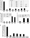
Isolation and characterization of BirA candidates exhibiting superrepressor phenotypes. Single colonies were streaked on defined medium plates supplemented with 1.6 nM biotin and X-Gal for phenotypic confirmation. Liquid cultures grown in the same medium were then assayed for bio operon transcription by β-galactosidase activity (Materials and Methods). The birA inserts of the plasmids encoding the superrepressor BirAs were then sequenced. Panel A shows the transcriptional activities and mutations of the mutants studied, whereas panel B illustrates the deconvolution of the double mutants 1A and 6A (which shared the I187T mutation) to identify the mutations causing the superrepressor phenotype. Panel C shows the deconvolution of the 12A triple mutant. The single mutations were generated by site-directed mutagenic repair of each mutant allele with the wild-type sequence. Strains carrying the plasmids encoding single mutant BirAs were then assayed for β-galactosidase activity. (D) Finally, the β-galactosidase activities of the plasmid-borne single mutant superrepressor strains chosen for in vivo and in vitro analyses were compared with the wild-type strain and with each other.
Fig 4
Fig 4
Complementation of the E. coli birA ΔbioABFCD strain by expression of either wild-type BirA (A) or G154D BirA (B) from a medium-copy-number plasmid. The strains were assayed for growth in defined medium liquid cultures supplemented with various concentrations of biotin as described in Materials and Methods. The different biotin concentrations are indicated by the colored symbols to the right of panel B. Each growth curve assay was carried out in triplicate, and the average was used in this plot. Growth was measured by optical density at 600 nm. Note that the y axis is plotted in a log scale. Panels A and B have the same color code.
Fig 5
Fig 5
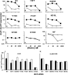
Regulation of bio operon expression in BirA mutant strains upon overexpression of biotin acceptor proteins. Shown is expression of the bio operon in cultures grown with various concentrations of biotin assayed by use of β-galactosidase production from the chromosomal bioF::lacZY fusion. The strains used were derivatives of strain VC618 cured of plasmid pVC17 that carried the lacIq plasmid pMS421 plus two compatible plasmids, one of which encoded a BirA mutant protein and the other of which encoded wild-type AccB plus AccC or K122R AccB-AccC or the biotin-accepting peptide 85 fusion protein. The AccB-AccC and K122R AccB-AccC plasmids were pCY705 and pCY730, respectively, whereas the plasmid pCY759 encoded the maltose binding protein fused to biotin-accepting peptide 85. The K122R AccB protein that is unable to accept biotin served as the control. The most responsive range of biotin concentrations (x axis) was 4 to 40 nM. Derivatives of strain VC618 carrying the wild-type or the mutant BirA plasmids plus pCY705 (■) or pCY730 (△) were induced with 0.1 mM IPTG. The experiments were repeated three times in their entirety, and the results were essentially identical to those shown. Also included are strain VC618 derivatives that carried the peptide 85 plasmid pCY759 and plasmids encoding either the G154D BirA mutant (H) or wild-type BirA (I). The symbol × represents assays with cultures that were induced with 0.1 mM IPTG, whereas ○ represents assays of uninduced cultures. Note that the y axis is plotted on a log scale. The data in panels J and K are from three independent experiments and demonstrate the effects of AccB overexpression on the regulation of the bio operon by the wild-type and birA mutant strains. The wild-type AccB-AccC expression (J) was performed at 40 nM biotin (repression conditions), whereas the K122R AccB-AccC (K) expression was assayed at 4 nM (derepression conditions). Open bars indicate cultures induced with 0.1 mM IPTG, whereas the solid bars indicate cultures that were not induced. Error bars denote the standard error. The strain expressing the G154D BirA mutant and pCY730 grew very poorly and therefore was unavailable for β-galactosidase assay (G and K).
Fig 6
Fig 6
Transcriptional regulation of the biotin operon mediated by the chromosomal birA alleles in cultures grown with various biotin concentrations. All strains contained the chromosomal bioF::lacZY fusion and originally carried plasmid pCY255 encoding yeast protein Bpl1. In panel A, the strains were grown in defined medium supplemented with various concentrations of biotin, as shown on the x axis. Gray bars denote wild-type birA allele, red bars denote the birA null (deletion [birAKO]) allele, and black bars denote the birA allele encoding G154D BirA. The results show the average of three independent experiments, and the error bars denote the standard error. Panel B shows the growth of strain VC780 (chromosomal wild-type BirA) or strain VC779 (chromosomal G154D BirA) or their polA12 derivatives (strains VC801 and VC802, respectively). The strains were streaked on defined medium plates supplemented with 4 nM biotin and X-Gal, which were incubated overnight at either 30°C or 42°C. The A and B designations of strains VC801 and VC802 denote two different transductants from the polA12 strain constructions. Strains VC780 and VC801 were derepressed (blue colonies) and grew at both temperatures due to the expression of wild-type BirA. Strain VC779 grew at both temperatures due to the presence of pCY255, whereas strain VC802 failed to grow at 42°C because pCY255 could no longer replicate due to loss of DNA polymerase I activity at the nonpermissive temperature, and strain VC779 grew at 42°C but remained repressed.
Fig 7
Fig 7
SDS-gel analysis of purified BirA and AccB-87 proteins. In panel A, the BirA proteins were subjected to electrophoresis in a 12% polyacrylamide gel, whereas in panel B, the gel used for AccB-87 was 15% polyacrylamide. G154D BirA consistently migrates more slowly than the other BirA proteins, a behavior that has been observed with other single-amino-acid substitutions (30).
Fig 8
Fig 8
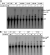
DNA binding properties of mutant BirA proteins assayed by EMSA. In panels A to D, the mutant and wild-type BirAs were assayed for binding to a 112-bp bioO duplex DNA labeled at the 5′ ends with 32P prepared as described in Materials and Methods. The reaction products were separated via native gel electrophoresis and visualized via phosphorimaging. The mutant and wild-type BirA proteins (0 to 500 nM) were assayed in parallel. The control reactions of all panels are indicated by “NE” (no enzyme added). From left to right, the BirA concentrations assayed were 500 nM, 250 nM, 125 nM, 61.5 nM, and 31.25 nM. Each panel contains the wild-type BirA plus a mutant BirA, as given. Panels A, B, C, and D, respectively, contained the G154D, K267M, I187T, and M310L BirA proteins.
Fig 9
Fig 9
Abilities of the mutant BirA proteins to synthesize biotinoyl-adenylate and transfer the biotin moiety to AccB-87. In panels A and B, synthesis of the biotinoyl-adenylate in reaction mixtures containing BirA (1 μM), biotin (0.01 μM), and [α-32P]ATP (0.1 μM) is shown. Following a 30-min incubation, each reaction mixture was split into halves, and to one-half (+) purified apo-AccB-87 (70 μM) was added followed by incubation for an additional 15 min, whereas the other half (−) were left untreated. One microliter of each sample was then spotted onto a cellulose thin-layer chromatography plate. Following plate development, the reaction products (indicated by arrows), biotinoyl-adenylate (bio-5′-AMP), and AMP were visualized by autoradiography. In panel B, similar experiments were carried out, except that two different concentrations of AccB-87 (7 μM or 70 μM) were used in the transfer assay. The control reactions of both panels are represented by “NB” (no biotin added) and “NE” (no enzyme added). The major spot on all lanes is ATP. + represents assays in which the acceptor AccB-87 was added, whereas − denotes assays in which the acceptor protein was not added.
Fig 10
Fig 10
The three-dimensional structure of BirA. In panel A, the three different domains of the monomeric protein are shown. The single-amino-acid substitutions that resulted in superrepressor phenotypes are highlighted. In panel B, the 2.8-Å crystal structure of dimeric BirA complexed to the corepressor analogue, biotinoyl-5′-AMP (colored white), is shown. Relative to the unliganded structure, corepressor analogue binding resulted in ordering of both adenylate-binding loops, and the dimer interface also undergoes a favorable transition that might be necessary for BirA binding to bioO. Several of the mutations are found within the dimer interface, suggesting that they might stabilize the dimer to give the observed superrepressor phenotypes. The model was generated by using the graphics program Chimera (32) with Protein Data Bank files 1HXD and 2EWN.

References

    1. Abdel-Hamid AM, Cronan JE. 2007. Coordinate expression of the acetyl coenzyme A carboxylase genes, accB and accC, is necessary for normal regulation of biotin synthesis in Escherichia coli. J. Bacteriol. 189:369–376 - PMC - PubMed
    1. Allison JL, et al. 1962. Mode of action of chloramphenicol. VII. Growth and multiplication of Escherichia coli in the presence of chloramphenicol. J. Bacteriol. 83:609–615 - PMC - PubMed
    1. Baba T, et al. 2006. Construction of Escherichia coli K-12 in-frame, single-gene knockout mutants: the Keio collection. Mol. Syst. Biol. 2:2006.0008 - PMC - PubMed
    1. Barker DF, Campbell AM. 1981. The birA gene of Escherichia coli encodes a biotin holoenzyme synthetase. J. Mol. Biol. 146:451–467 - PubMed
    1. Barker DF, Campbell AM. 1981. Genetic and biochemical characterization of the birA gene and its product: evidence for a direct role of biotin holoenzyme synthetase in repression of the biotin operon in Escherichia coli. J. Mol. Biol. 146:469–492 - PubMed

Publication types

MeSH terms

LinkOut - more resources