. 2012 Mar;40(6):e45.
doi: 10.1093/nar/gkr1262. Epub 2011 Dec 30.

In vitro experimental system for analysis of transcription-translation coupling

Affiliations

In vitro experimental system for analysis of transcription-translation coupling

Daniel Castro-Roa et al. Nucleic Acids Res. 2012 Mar.

Abstract

Transcription and translation are coupled in bacteria, meaning that translation takes place co-transcriptionally. During transcription-translation, both machineries mutually affect each others' functions, which is important for regulation of gene expression. Analysis of interactions between RNA polymerase (RNAP) and the ribosome, however, are limited due to the lack of an in vitro experimental system. Here, we report the development of an in vitro transcription coupled to translation system assembled from purified components. The system allows controlled stepwise transcription and simultaneous stepwise translation of the nascent RNA, and permits investigation of the interactions of RNAP with the ribosome, as well as the effects of translation on transcription and transcription on translation. As an example of usage of this experimental system, we uncover complex effects of transcription-translation coupling on pausing of transcription.


Figures

Figure 1.
Figure 1.
In vitro characterization of the translational machinery. (A) Schematic representation of the toeprinting of ribosome front edge and RelE cleavage site. Reverse transcription of mRNA (red line) results in synthesis of 5′-end labeled cDNA (blue line) of either full length in the absence of ribosomes (beige circle), and shorter cDNAs corresponding to stops of reverse transcriptase (green circle) in front of ribosomes or at the site of RelE (blue triangle) cleavage. Elongation by ribosome to specific positions (MF and MFV) towards the 3′-end of the RNA results in formation of shorter cDNA. RelE cleavage at the vacant A site of ribosome (dark beige half) results in dissociation of the ribosome and synthesis of cDNA to the cleavage site. (B) Toeprinting of initiating and translocating ribosomes on gene 32 mRNA. Shown is denaturing PAGE of toeprint cDNA's of ribosomes initiated in factor-independent (lanes 2–4, 8–9) or enzymatic (lanes 5–7, 10–11) manner and subsequently translocated in EF-G dependent (lanes 3, 4, 6, 7) and EF-G independent (8–11) manner to positions designated to the left of the gel. (C) Denaturing PAGE of cDNA's forming during titration of RelE (1, 4, 8, 14 pmol in lanes 4, 5, 6, 7, respectively) on translation initiation complex. Note that the lower bands present in lanes 1 and 2 are due to inhibition of reverse transcription by the secondary structure in the mRNA. This secondary structure melts when the ribosome is loaded onto the mRNA.
Figure 2.
Figure 2.
Characterization of promoter borne CTT. (A) Schematic representation of promoter borne CTT system assembly with non-enzymatic translation initiation. Biotinylated DNA template (for full sequence see Supplementary Data) containing T7A1 promoter utilized by E. coli is immobilized on streptavidin beads. Escherichia coli RNAP initiated from the promoter is ‘walked’ to a desired position. Translation initiation and elongation complexes formed on the mRNA can be analyzed by RelE cleavage. (B) Example of walking of RNAP. PAGE of the RNAs synthesized by RNAP during its walking to position +80 (to form EC80). (C) RelE mapping (for times designated above the gel) of translation initiation complexes in CTT system assembled with promoter borne transcription EC80 (lanes 4–5). EC80 was obtained as in panel B, and the transcript was radiolabeled at its 5′-end proximal part. RNAs from transcription ECs stalled at positions +47, +48 and +51 (lanes 1–3) were used as size markers to determine the size of the RelE cleavage product. Note that degradation of EC80 is caused not only by RelE cleavage but also by RNAP-dependent phosphorolysis by high phosphate of RelE storage buffer. (D) PAGE of radiolabeled mRNA of CTT (assembled with stalled EC80), in which translation initiation (lanes 2 and 6) and elongation (in the presence of EF-G; lanes 3 and 7) are probed by RelE cleavage. In lanes 5–7, EC80 was chased in the presence of all NTPs after translation initiation and elongation but before RelE cleavage. EC80 was obtained as in panel B, and the transcript was radiolabeled at its 5′-end proximal part. RelE cleavage products were identified as in panel C by walking RNAP to the corresponding positions and loading these RNAs as markers. A weaker band of RelE cleavage product after ribosome translocation is explained by different activity of RelE on various codons. Note, some transcription read through from EC80 (lanes 3, 4) in the presence of GTP required for translocation. Black vertical lines separate lanes originating from one gel which were brought together.
Figure 3.
Figure 3.
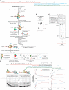
Characterization of ‘translation first’ TL-CTT. (A) Schematic representation of assembly of TL-CTT. Translation is initiated with f-Met-tRNAfmet (green triangle) and initiation factors 1, 2 and 3 (green, blue and red circles). After translation initiation, ribosomes are allowed to elongate by one codon with synthesis of a dipeptide. The translation elongation complexes are separated from unused factors and unused mRNA by ultracentrifugation through a sucrose cushion. The mRNA carrying elongating ribosome is used in assembly of transcription EC. mRNA in TL-CTT is labeled by incorporation of radiolabeled NMP during RNAP walking, ensuring that mRNA is labeled only in coupled complexes. Sequences of the nucleic acids scaffold used for transcription EC assembly are shown at the top of the panel. Nucleotides incorporated after CTT assembly are in black, asterisk represents the radiolabeled nucleotide. The ribosome can then be translocated allowing collisions to occur between the two machineries, followed by the analysis of transcripts by denaturing PAGE. (B) Peptidyl transferase assay to analyze the activity of the ribosome after purification and TL-CTT assembly. MF dipeptide (formed prior purification with [35S]-f-Met-tRNAfmet; lane 1) was allowed to be extended to tetrapeptide (MFVV; lane 2). Products were resolved by thin layer electrophoresis. (C) Occupancy of mRNAs with ribosomes in TL-CTT revealed by RelE cleavage. Shown is PAGE of mRNA. Note that only coupled complexes are visible since mRNA is labeled during RNAP transcription after TL-CTT assembly. RelE cleavage was performed in the translation elongation complex containing dipeptide MF (formed prior purification) and after this complex was allowed to elongate by two codons (to tetrapeptide MFVV). The sequence below shows where RelE cleavage takes place. (D) Complex effects of coupling on pausing of transcription as an example of using TL-CTT. Shown is PAGE of mRNA. Sequences of oligonucleotides used for TL-CTT assembly are shown at the top of the panel. Nucleotides incorporated after CTT assembly are in black, asterisk represents the radiolabeled nucleotide, arrows show pauses P1, P2, P3 and P4 formed during chase (positions of pauses were determined by walking RNAP to each position; not shown). After TL-CTT formation, RNAP was walked to P1 by addition of incomplete set of NTP's. Then, translation elongation complex was either left with dipeptide MF (‘stalled’ complexes) or was allowed to elongate behind RNAP by addition of F and V ternary complexes in presence of EF-G and GTP for 3 min (‘translating’ complexes). After this step, RNAP was allowed to transcribe by addition of four NTPs. While the stalled ribosomes remain at their initial position, the translating ribosomes follow transcribing RNAP. Gel shows effects on transcriptional pausing by coupled ribosome. Plots show quantification of some pauses as a fraction (in percent) of all complexes in the lane versus time. Error bars are standard deviation from two independent experiments.
Figure 4.
Figure 4.
Characterization of ‘transcription first’ TR-CTT. (A) Schematic representation of assembly of TR-CTT. Solid phase immobilized transcription EC (for full scaffold sequence see Supplementary Data) is washed to remove unincorporated mRNA, after that translation is initiated and allowed to elongate on the mRNA of the EC. (B) Peptidyl transferase activity in TR-CTT. Ribosome elongation by F, FV or FVY codons is followed by thin layer electrophoresis (TLE) of synthesized peptides labeled with [35S]-f-Met-tRNAfmet. Comparison of the peptidyl transferase activity of the ribosome on naked (without transcription EC) mRNA (lanes 1–3) and in the TR-CTT (lanes 4–6). (C) Not all transcription ECs are coupled to translation in TR-CTT as evidenced by RelE cleavage after TR-CTT assembly. Shown is PAGE of mRNA labeled during RNAP walking as in Figure 3. (D) Rough estimation of mRNA length behind transcribing RNAP sufficient for translation initiation as an example of using TR-CTT. MF dipeptide formation was used as a measure of translation initiation efficiency (given that it does not require translocation) on mRNAs containing 31- and 42-nt spacers between its 3′-end occupied by RNAP and the AUG start codon.

References

    1. Pomerantz RT, O'Donnell M. The replisome uses mRNA as a primer after colliding with RNA polymerase. Nature. 2008;456:762–766. - PMC - PubMed
    1. Zenkin N, Naryshkina T, Kuznedelov K, Severinov K. The mechanism of DNA replication primer synthesis by RNA polymerase. Nature. 2006;439:617–620. - PubMed
    1. Mirkin EV, Castro Roa D, Nudler E, Mirkin SM. Transcription regulatory elements are punctuation marks for DNA replication. Proc. Natl Acad. Sci. USA. 2006;103:7276–7281. - PMC - PubMed
    1. Azvolinsky A, Giresi PG, Lieb JD, Zakian VA. Highly transcribed RNA polymerase II genes are impediments to replication fork progression in Saccharomyces cerevisiae. Mol. Cell. 2009;34:722–734. - PMC - PubMed
    1. Merrikh H, Machón C, Grainger WH, Grossman AD, Soultanas P. Co-directional replication-transcription conflicts lead to replication restart. Nature. 2011;470:554–557. - PMC - PubMed

Publication types

MeSH terms

Substances