. 2012 Apr;40(8):3484-95.
doi: 10.1093/nar/gkr1245. Epub 2011 Dec 30.

Role of polynucleotide kinase/phosphatase in mitochondrial DNA repair

Affiliations

Role of polynucleotide kinase/phosphatase in mitochondrial DNA repair

Nasser Tahbaz et al. Nucleic Acids Res. 2012 Apr.

Abstract

Mutations in mitochondrial DNA (mtDNA) are implicated in a broad range of human diseases and in aging. Compared to nuclear DNA, mtDNA is more highly exposed to oxidative damage due to its proximity to the respiratory chain and the lack of protection afforded by chromatin-associated proteins. While repair of oxidative damage to the bases in mtDNA through the base excision repair pathway has been well studied, the repair of oxidatively induced strand breaks in mtDNA has been less thoroughly examined. Polynucleotide kinase/phosphatase (PNKP) processes strand-break termini to render them chemically compatible for the subsequent action of DNA polymerases and ligases. Here, we demonstrate that functionally active full-length PNKP is present in mitochondria as well as nuclei. Downregulation of PNKP results in an accumulation of strand breaks in mtDNA of hydrogen peroxide-treated cells. Full restoration of repair of the H(2)O(2)-induced strand breaks in mitochondria requires both the kinase and phosphatase activities of PNKP. We also demonstrate that PNKP contains a mitochondrial-targeting signal close to the C-terminus of the protein. We further show that PNKP associates with the mitochondrial protein mitofilin. Interaction with mitofilin may serve to translocate PNKP into mitochondria.


Figures
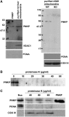
Figure 1.
Figure 1.
Full-length PNKP is present in mitochondria. (A) Mitochondria were isolated from rat liver and wild-type (WT) and PNKP knock-down (KD) A549 cells. Mitochondrial protein extracts were immunoblotted with a monoclonal antibody to PNKP. Antibodies against PCNA (nuclear marker) and COX IV or VDAC1 (mitochondrial markers) were used to ensure the purity of the mitochondrial preparation. (B) Purified human PNKP protein is sensitive to proteinase K, and is completely digested even at the lowest concentration used (10 µg ml−1). (C) PNKP signal detected in purified mitochondria isolated from A549 cells is protected from proteinase K digestion. Proteins in nuclear extracts are shown on the left and purified PNKP protein on the right.
Figure 2.
Figure 2.
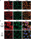
Endogenous PNKP colocalizes with mitochondrial markers. (A) Immunofluorescence of PNKP in A549 cells. PNKP localizes to mitochondria (in addition to the nuclei) as demonstrated by colocalization with the mitochondrial markers mitofilin and COX IV. (B) The shRNA KD of PNKP (lower panel) results in a decrease of the fluorescence signal for PNKP in both nuclei and mitochondria of A549 cells compared to the control cell line carrying scrambled shRNA (upper pannel).
Figure 3.
Figure 3.
Mitochondrial PNKP displays both DNA kinase and phosphatase activities. (A) DNA kinase activity was measured by transfer of 32P-phosphate from ATP to an oligonucleotide (21-mer) with a 5′-hydroxyl terminus. Mitochondria were purified from wild-type (WT) and PNKP KD A549 cells and treated with trypsin to digest any possible residual extramitochondrial protein contamination. Trypsin-treated mitochondria, either sonicated (+) or not sonicated (−), were used for DNA kinase assays. Sonication increased the DNA kinase activity of the purified mitochondrial preparation in WT cells. DNA kinase activity of T4 PNK is shown as a positive control. (B) DNA 3′-phosphatase activity in mitochondrial preparations from WT and KD A549 cells was determined by dephosphorylation of a 5′-32P-labelled 3′-phosphorylated oligonucleotide (p20p), as substrate, resulting in conversion of p20p to p20 (markers shown on left). Sonication of the purified mitochondrial preparation substantially increased DNA phosphatase activity in WT cells.
Figure 4.
Figure 4.
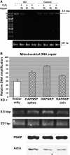
Functional PNKP is required for DNA repair in mitochondria following exposure to H2O2. (A) Total (i.e. nuclear + mitochondrial) DNA was purified from A549 cell lines stably transfected with PNKP shRNA (KD) or scrambled shRNA (control) (Supplementary Figure S3) following exposure to 1.5 mM H2O2 for 1 h and repair for 0, 2 and 4 h. Controls (−) were not exposed to H2O2. Upper panel: XL-qPCR performed on mtDNA, as described in Materials and Methods, amplified an 8.9-kbp PCR product following H2O2 exposure and repair in A549 cells expressing scrambled shRNA, but not in PNKP KD A549 cells. Lower panel: PCR of a 221-bp fragment from both cell lines indicating that both lines contain comparable amounts of mtDNA and that the H2O2 treatment did not degrade the DNA. (B) DNA repair in mitochondria in PNKP KD cells complemented with empty vector, wild-type (HAPNKP) or kinase (HAPNKPΔkin) or phosphatase inactive (HAPNKPΔphos) PNKP. XL-qPCR was used to examine the level of DNA repair 30 min after exposure to 1 mM H2O2 for 1 h. The signal from the amplified 8.9 kbp mtDNA following XL-qPCR was normalized to the signal from the 221-bp fragment using Quant-it Pico Green Assay Kit. Error bars show the SD for three independent experiments. The western blot shows that similar levels of PNKP protein were expressed in the PNKP KD background.
Figure 5.
Figure 5.
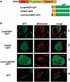
Mitochondrial localization of PNKP is dependent on the presence of a mitochondrial-targeting signal (MTS) in proximity to its carboxy terminus. (A) Computer programs (Mitoprot, Psort II and Predotar) predicted the presence of a mitochondrial-targeting signal (MTS) close to the C-terminus of PNKP (shown in blue). To further examine this potential MTS we generated three constructs (i) CmtsPNKP + GFP containing the GFP fused to the PNKP C-terminus including the putative MTS, (ii) CPNKP + GFP, which is essentially the same as CmtsPNKP + GFP but lacking the MTS sequence and (iii) mutCmtsPNKP + GFP, a mutated form of CmtsPNKP + GFP with the first three amino acids of the putative PNKP MTS mutated as follows: A432D, R433G and Y434D. In all cases a methionine was included at the amino-terminus. (B) The constructs were transfected into A549 cells and the cellular localization of GFP was monitored. Only the GFP fusion protein containing the wild-type MTS localized to mitochondria (row 1) as shown by colocalization with Mitotracker Orange (Molecular Probes). CPNKP + GFP, mutCmtsPNKP + GFP and GFP alone showed a diffuse signal throughout the cell (rows 2–4).
Figure 6.
Figure 6.
The MTS of PNKP is required for its function in mtDNA repair. XL-qPCR was used to monitor mtDNA repair in PNKP-depleted A549 cells treated with hydrogen peroxide as described in Figure 4B. Transient complementation of the cells with PNKP mutated in the first three amino acids of the MTS (HAPNKP-mts), as opposed to the wild-type protein (HAPNKP), reduces DNA repair in mitochondria to a level similar to the vector only control. The western blot of whole-cell extracts shown at the bottom of the figure indicates that similar levels of HAPNKP-mts and HAPNKP proteins were expressed in the A549 cells.
Figure 7.
Figure 7.
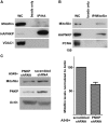
PNKP physically interacts with mitochondrial proteins. (A) Coimmunopreciptation of mitofilin by anti-HA antibody from whole-cell extract (WC) of A549 cells expressing HAPNKP. An antibody to the mitochondrial protein VDAC1 was used to ensure no non-specific pull-down of mitochondrial proteins. (B) Coimmunoprecipitation of PNKP with mitofilin from whole-cell extract of A549 cells expressing HAPNKP using a monoclonal antibody to mitofilin to immunoprecipitate mitofilin. In this case, the immunopreciptate was also tested for the presence of PCNA as a marker of potential contamination by nuclear proteins. The control lane ‘beads only’ indicates that no PNKP bound to the Sepharose beads in the absence of antibodies. (C) KD of PNKP by shRNA results in a decrease of about 40% in cellular mitofilin content. The graph shows the mean and SD values from three independent determinations (P = 0.013).

Similar articles

Cited by

References

    1. Henner WD, Grunberg SM, Haseltine WA. Sites and structure of gamma radiation-induced DNA strand breaks. J. Biol. Chem. 1982;257:11750–11754. - PubMed
    1. Lennartz M, Coquerelle T, Bopp A, Hagen U. Oxygen—effect on strand breaks and specific end-groups in DNA of irradiated thymocytes. Int. J. Radiat. Biol. Relat. Stud. Phys. Chem. Med. 1975;27:577–587. - PubMed
    1. Weinfeld M, Mani RS, Abdou I, Aceytuno RD, Glover JN. Tidying up loose ends: the role of polynucleotide kinase/phosphatase in DNA strand break repair. Trends Biochem. Sci. 2011;36:262–271. - PMC - PubMed
    1. Pourquier P, Pilon AA, Kohlhagen G, Mazumder A, Sharma A, Pommier Y. Trapping of mammalian topoisomerase I and recombinations induced by damaged DNA containing nicks or gaps. Importance of DNA end phosphorylation and camptothecin effects. J. Biol. Chem. 1997;272:26441–26447. - PubMed
    1. Plo I, Liao ZY, Barcelo JM, Kohlhagen G, Caldecott KW, Weinfeld M, Pommier Y. Association of XRCC1 and tyrosyl DNA phosphodiesterase (Tdp1) for the repair of topoisomerase I-mediated DNA lesions. DNA Repair. 2003;2:1087–1100. - PubMed

Publication types

Substances