. 2012 May;40(9):4146-57.
doi: 10.1093/nar/gkr1281. Epub 2011 Dec 30.

Crystal structure of human polynucleotide phosphorylase: insights into its domain function in RNA binding and degradation

Affiliations

Crystal structure of human polynucleotide phosphorylase: insights into its domain function in RNA binding and degradation

Chia Liang Lin et al. Nucleic Acids Res. 2012 May.

Abstract

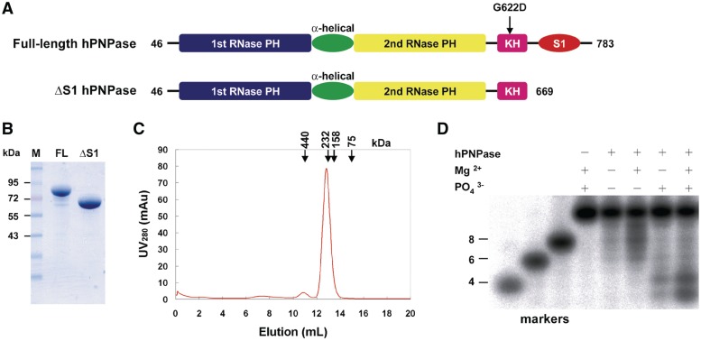
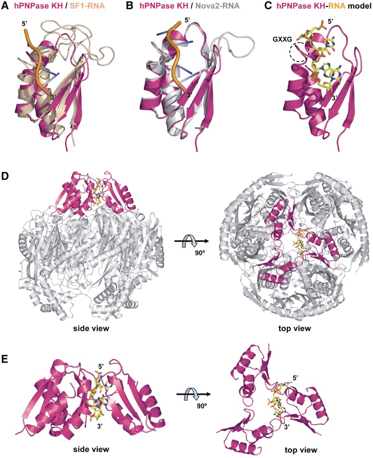
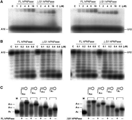
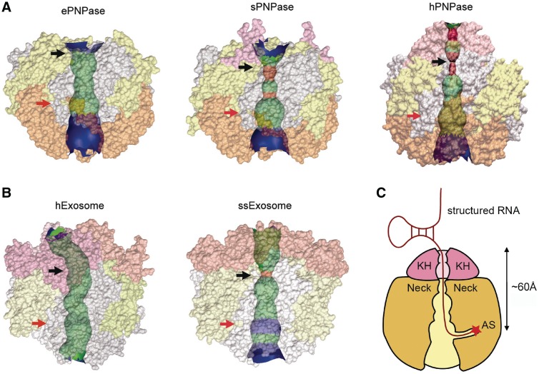
Human polynucleotide phosphorylase (hPNPase) is a 3'-to-5' exoribonuclease that degrades specific mRNA and miRNA, and imports RNA into mitochondria, and thus regulates diverse physiological processes, including cellular senescence and homeostasis. However, the RNA-processing mechanism by hPNPase, particularly how RNA is bound via its various domains, remains obscure. Here, we report the crystal structure of an S1 domain-truncated hPNPase at a resolution of 2.1 Å. The trimeric hPNPase has a hexameric ring-like structure formed by six RNase PH domains, capped with a trimeric KH pore. Our biochemical and mutagenesis studies suggest that the S1 domain is not critical for RNA binding, and conversely, that the conserved GXXG motif in the KH domain directly participates in RNA binding in hPNPase. Our studies thus provide structural and functional insights into hPNPase, which uses a KH pore to trap a long RNA 3' tail that is further delivered into an RNase PH channel for the degradation process. Structural RNA with short 3' tails are, on the other hand, transported but not digested by hPNPase.


Figures

Figure 1.
Figure 1.
Recombinant human PNPase is a trimeric phosphorylase capable of digesting RNA. (A) Domain organization of full-length (FL) and S1 domain-truncated (ΔS1) hPNPase. (B) Purity of full-length and ΔS1 hPNPase, as analyzed by 10% SDS–PAGE. (C) The gel filtration (Superdex 200) profile of full-length hPNPase shows that the enzyme was eluted as a trimeric protein. Protein markers: ferritin (440 kDa), catalase (232 kDa), aldolase (158 kDa) and conalbumin (75 kDa). (D) The full-length hPNPase (0.2 µM) was incubated for 30 min at 37°C with isotope-labeled 12-mer poly(A) ssRNA. In the presence of both magnesium and phosphate ions, hPNPase cleaved RNA most efficiently.
Figure 2.
Figure 2.
Crystal structure of ΔS1 hPNPase. (A) The monomeric structure of ΔS1 hPNPase comprises two RNase PH domains (blue and yellow), an α-helical domain (green) and a KH domain (magenta). Two citrate ions (orange) bound near the active site are displayed in stick models. (B) Side view and top view of the trimeric ΔS1 hPNPase which assembled into a ring-like structure with a central channel. The three KH domains are located on the top of the ring, forming a novel RNA-binding KH pore. (C) Superimposition of the structure of ΔS1 hPNPase (PDB entry 3U1K, magenta) with those of S. antibioticus PNPase (PDB entry 1E3P, yellow) and E. coli PNPase (PDB entry 3CDI, gray). The RNase PH and α-helical domains matched well; however, the KH domain in hPNPase (marked by a dashed black oval) does not superimpose with the poly-alanine chain in S. antibioticus PNPase (marked by a dashed red oval). (D) Two citrate ions (stick models in orange) bind at the active site in the second RNase PH domain in hPNPase. (E) Trimeric and monomeric full-length structural models of hPNPase generated by superimposition of the structure of ΔS1 hPNPase (PDB entry 3U1K) with those of S. antibioticus PNPase (PDB entry 1E3P) and S1 domain of E. coli PNPase (PDB entry 1SRO). Blue, magenta and yellow represent S1 domain, KH domain and RNase PH domain, respectively.
Figure 3.
Figure 3.
Comparison of the S1 pore in exosomes and the KH pore in hPNPase. (A) The archaeal exosome (PDB entry 2JE6) has an S1 pore formed by the three S1 domains of Rrp4 (blue). The human exosome (PDB entry 2NN6) has a more open S1 pore formed by the S1 domains of Rrp4, Rrp40 and Csl4 (blue). In contrast, human PNPase has a KH pore formed by three KH domains (magenta). (B) Molecular surface representation of the archaeal exosome, human exosome and human PNPase structures show clearly differences in the arrangement of the domains that form the S1 pore and KH pore in exosomes and hPNPase, respectively. The color code is the same as in panel A: S1 domain in blue, KH domain in magenta, RNase PH domain in yellow, NTD domain (in Rrp40, Rrp4 and Csl4) in gray and CTD domain (in Csl4) in beige.
Figure 4.
Figure 4.
Structural model of RNA bound at the KH pore of hPNPase. (A) Superimposition of the hPNPase KH domain (magenta) with the KH domain in the SF1–RNA complex structure (PDB entry 1K1G, beige for SF1 and orange for RNA) gave an average RMSD of 3.12 Å for 30 Cα atoms. (B) Superimposition of the hPNPase KH domain (magenta) with the KH domain in the Nova2–RNA complex structure (PDB entry 1EC6, gray for Nova2 and orange for RNA) gave an average RMSD of 1.45 Å for 48 Cα atoms. (C) Structural model of the KH domain (magenta) of hPNPase in complex with a single-stranded, 4-nt RNA adapted from Nova2-RNA. The GXXG motif is marked by a dashed circle. (D) Structural model of the hPNPase–RNA complex. The 4-nt RNA is bound inside the KH pore. (E) A close-up view of the trimeric KH pore in hPNPase bound with RNA. The 3′-end of the RNA points inward into the channel.
Figure 5.
Figure 5.
RNA binding and cleavage assays for full-length (FL) and ΔS1 hPNPase. (A) EMSA assay showing that the RNA substrate binds with a similar affinity to full-length and ΔS1 hPNPase. The 12-mer poly(A) and poly(U) ssRNA substrate (0.1 pmol) was incubated, respectively, with hPNPase at various concentrations ranging from 1 to 16 µM in the absence of phosphate and Mg2+ ions. (B) The RNase activity of full-length and ΔS1 hPNPase was examined by incubation of the ssRNA with different concentrations of protein (0.1–0.8 µM) for 30 min at 37°C. Full-length and ΔS1 hPNPase exhibited a similar RNase activity. (C) Full-length and ΔS1 hPNPase can digest the long 3′ overhang (15 and 20 nt) of stem–loop RNA and generate major products with an overhang of 11–14 nt. The stem–loop RNA with a short 3′ overhang (7 and 10 nt) were more resistant for digestion. The marker (M) of 20-nt RNA corresponds to the stem–loop region of RNA with an 8-bp duplex and a 4-nt loop. The A0, A10 and A15 indicate the overhang size of 0, 10 and 15 nt, respectively.
Figure 6.
Figure 6.
The GXXG motif in the KH domain of PNPase participates in RNA binding. (A) Sequence alignment of the KH domain of PNPase from different species shows that the GXXG motif is highly conserved. Secondary structure elements are shown on the top, with arrows indicating β-sheets and cylinders indicating α-helices. The PNPase-specific GXXG sequences are marked with a box. Sequences listed here include PNPase from Homo sapiens (Hs), Arabidopsis thaliana (At), Spinacia oleracea (So), E. coli (Ec) and S. antibioticus (Sa). (B) Sequence alignment of the KH domain in Rrp40 and Rrp4 exosome components shows that the GXXG motif was not conserved. Sequences listed here include components from the following species: Saccharomyces cerevisiae (Sc), H. sapiens (Hs), S. solfataricus (Ss) and Archaeoglobus fulgidus (Af). (C) EMSA assay showing that the hPNPase mutant G622D lost its RNA-binding activity in binding a 12-mer poly(A) ssRNA. (D) RNase activity assay showing that the hPNPase mutant G622D digested the 12-mer poly(A) RNA poorly as compared to wild-type PNPase.
Figure 7.
Figure 7.
The RNA-binding central channel in PNPase and exosomes. (A) Comparison between the central channel in E. coli PNPase (ePNPase, PDB: 3CDI), S. antibioticus PNPase (sPNPase, PDB: 1E3P) and hPNPase (PDB: 3U1K) shows that hPNPase has the most constricted channel for RNA binding among these PNPases [calculated by using the program HOLE (47)]. The channel regions with a diameter of <5 Å are displayed in red and pink. The neck regions are marked by black arrows and the active sites are marked by red arrows. (B) The crystal structures of human exosome (PDB: 2NN6) and S. solfataricus exosome (PDB: 2JE6) show that the active S. solfataricus exosome has a more constricted channel as compared to the inactive human exosome. (C) Structural model of hPNPase bound with a structured RNA.

Similar articles

Cited by

References

    1. Mohanty BK, Kushner SR. Polynucleotide phosphorylase functions both as a 3′→5′ exonuclease and a poly(A) polymerase in Escherichia coli. Proc. Natl Acad. Sci. USA. 2000;97:11966–11971. - PMC - PubMed
    1. Deutscher MP. Degradation of RNA in bacteria: comparison of mRNA and stable RNA. Nucleic Acids Res. 2006;34:659–666. - PMC - PubMed
    1. Andrade JM, Pobre V, Silva IJ, Domingues S, Arraiano CM. The role of 3′–5′ exoribonucleases in RNA degradation. Prog. Mol. Biol. Trans. Sci. 2009;85:187–229. - PubMed
    1. Das SK, Bhutia SK, Stokhi UK, Dash R, Azab B, Sarkar D, Fisher PB. Human polynucleotide phosphorylase (hPNPaseold-35): an evolutionary conserved gene with an expanding repertoire of RNA degradation functions. Oncogene. 2011;30:1733–1743. - PMC - PubMed
    1. Marcaida MJ, DePristo MA, Chandran V, Carpousis AJ, Luisi BF. The RNA degradosome: life in the fast lane of adaptive molecular evolution. Trends Biochem. Sci. 2006;31:359–365. - PubMed

Publication types

MeSH terms

Associated data