. 2012 Feb 1;188(3):992-1001.
doi: 10.4049/jimmunol.1102098. Epub 2011 Dec 30.

TLR2 activation enhances HIV nuclear import and infection through T cell activation-independent and -dependent pathways

Affiliations

TLR2 activation enhances HIV nuclear import and infection through T cell activation-independent and -dependent pathways

Jian Ding et al. J Immunol. .

Abstract

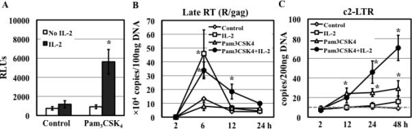
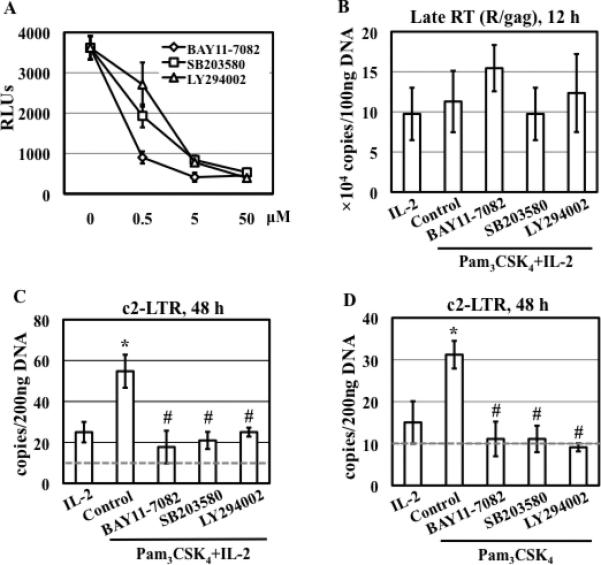
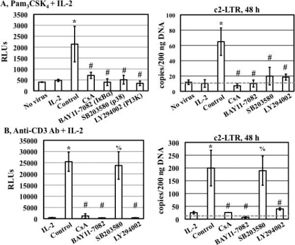
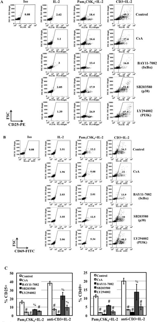
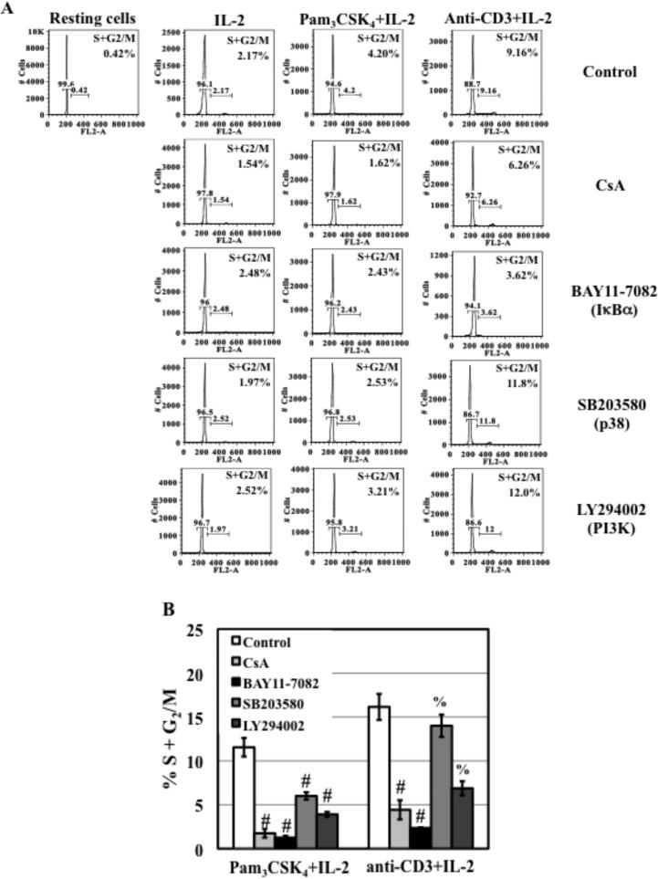
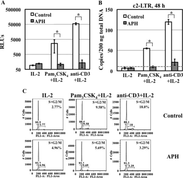
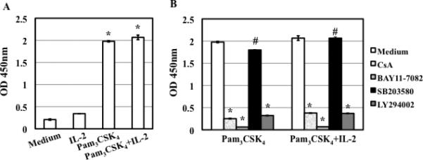
TLR2 activation plays a crucial role in Neisseria gonorrheae-mediated enhancement of HIV infection of resting CD4(+) T cells. We examined signaling pathways involved in the HIV enhancing effect of TLR2. TLR2 but not IL-2 signals promoted HIV nuclear import; however, both signals were required for the maximal enhancing effect. Although TLR2 signaling could not activate T cells, it increased IL-2-induced T cell activation. Cyclosporin A and IkBα inhibitor blocked TLR2-mediated enhancement of HIV infection/nuclear import. PI3K inhibitor blocked HIV infection/nuclear import and T cell activation and exerted a moderate inhibitory effect on cell cycle progression in CD4(+) T cells activated by TLR2/IL-2. Blockade of p38 signaling suppressed TLR2-mediated enhancement of HIV nuclear import/infection. However, the p38 inhibitor did not have a significant effect on T cell activation or TCR/CD3-mediated enhancement of HIV infection/nuclear import. The cell cycle arresting reagent aphidicolin blocked TLR2- and TCR/CD3-induced HIV infection/nuclear import. Finally, cyclosporin A and IκBα and PI3K inhibitors but not the p38 inhibitor blocked TLR2-mediated IκBα phosphorylation. Our results suggest that TLR2 activation enhances HIV infection/nuclear import in resting CD4(+) T cells through both T cell activation-dependent and -independent mechanisms.


Figures

Figure 1
Figure 1. IL-2 is required for the maximal HIV enhancing effect of TLR2 activation
(A) Primary resting CD4+ T cells were infected with pseudotyped HIV-1VSV-G luciferase reporter virus and then treated with medium (control) or Pam3CSK4 (5 μg/mL) in the presence or absence of IL-2. HIV infection was determined by measuring luciferase activity at day 4 after infection. The difference between samples treated with Pam3CSK4 in the presence or absence of IL-2 is significant (*P<0.05). (B-C) HIV-infected resting CD4+ T cells were treated with medium (control) or Pam3CSK4 in the presence or absence of IL-2. Total DNA was prepared at indicated time points after infection. The levels of HIV-1 late RT (R/gag) products (B) and c2-LTR circles (C) were determined by quantitative real-time PCR analysis. The detection limit (gray dashed line in panel C) for c2-LTR circles was 10 copies per 200 ng total DNA. There is a difference in the level of RT products between control samples and those treated with IL-2 alone or with Pam3CSK4 and IL-2 (*P<0.05). The difference in the level of c2-LTR circles between control samples and those treated with Pam3CSK4 or with Pam3CSK4 and IL-2 is significant. Results presented are means ± SD of triplicate sample and represent 3 independent experiments.
Figure 2
Figure 2. Kinase specific inhibitors block TLR2-mediated enhancement of HIV infection and nuclear import in resting CD4+ T cells
(A) HIV-infected primary resting CD4+ T cells were treated with kinase specific inhibitors at various concentrations for 1 h before stimulation with Pam3CSK4 and IL-2 (in the continued presence of inhibitors). HIV infection was determined by measuring luciferase activity at day 4 after infection. (B) HIV-infected resting CD4+ T cells were treated with inhibitors (15 μM) for 1 h and then stimulated with Pam3CSK4 and IL-2. The level of HIV-1 late RT products was analyzed 12 h after infection. There is no difference in the levels of late RT products between samples with or without exposure to inhibitors (P>0.05). (C) HIV-infected resting CD4+ T cells were treated with inhibitors (15 μM) for 1 h followed by stimulation with Pam3CSK4 and IL-2 for 48 h. The level of c2-LTR circles was measured by real-time PCR analysis. There is a significant difference between samples with treatment of IL-2 and of Pam3CSK4/IL-2 (*P<0.05) as well as between cells with treatment of Pam3CSK4 /IL-2 in the presence or absence of inhibitors (#P<0.05). (D) HIV-infected resting CD4+ T cells were treated with inhibitors (15 μM) for 1 h followed by stimulation with Pam3CSK4 in the absence of IL-2 for 48 h. The level of c2-LTR circles was determined. There is a significant difference between samples treated with Pam3CSK4 and those in media alone or in the presence of IL-2 (*P<0.05). There is a difference between samples with treatment of Pam3CSK4 in the presence or absence of inhibitors (#P<0.05). Data presented are means ± SD of triplicate sample and represent 3 independent experiments.
Figure 3
Figure 3. p38 inhibitor SB203580 blocks enhancement of HIV infection and nuclear import in resting CD4+ T cells in response to TLR2 but not TCR/CD3 activation
HIV-infected primary resting CD4+ T cells were treated with medium, CsA (1 μg/mL) and specific kinase inhibitors (15 μM) for 1 h before stimulation with either Pam3CSK4 (A) or anti-CD3 Ab at 1 μg/mL (B) in the presence of IL-2. Luciferase activity was measured at day 4 after infection, and the level of c2-LTR circles was determined at 48 h after infection. There is a significant difference between samples with treatment of IL-2 alone or those with Pam3CSK4/IL-2 or anti-CD3 Ab/IL-2 (*P<0.05). The difference between Pam3CSK4/IL-2-treated cells in the presence or absence of inhibitors is significant (#P<0.05). The difference between anti-CD3/IL-2-treated cells in the presence or absence of CsA, BAY11-7082, and LY294002, but not SB203580, is significant (#P<0.05; %P>0.05). The results shown are means ± SD of triplicate samples and are representative of results from 3 donors.
Figure 4
Figure 4. The effect of CsA and kinase specific inhibitors on T cell activation in response to TLR2 and TCR/CD3 activation
(A and B) Primary resting CD4+ T cells were treated with CsA (1 μg/mL) and kinase specific inhibitors (15 μM) before stimulation with Pam3CSK4 (5 μg/mL)/IL-2 or anti-CD3 Ab (1 μg/mL)/IL-2 for 3 days before analysis of surface expression of CD25 and CD69. Results from other donors were shown in Table S1. (C) The effect of inhibitors at higher concentrations (CsA at 10 μg/mL and kinase specific inhibitors at 50 μM) on T cell activation was determined. Data are presented as the means ± SD of the results from 3 donors. The difference in CD25 and CD69 expression between activated cells in the presence or absence of inhibitors is indicated (#P<0.05) as determined by one tail, paired Student's t test. Compared to the samples in the absence of inhibitors, the p-values for TLR2-treated samples in the presence of the p38 inhibitor SB203580 and PI3K inhibitor LY294002 were 0.068 and 0.03, respectively. There is no significant difference for CD25 and CD69 expression between anti-CD3-activated cells in the presence or absence of SB203580 (%P>0.05, p=0.15 and 0.2 for CD25 and CD69, respectively).
Figure 5
Figure 5. The effect of CsA and kinase inhibitors on cell cycle progression from G0/G1 to S and G2/M phases in response to TLR2 and TCR/CD3 activation
(A) Primary resting CD4+ T cells were treated with CsA (1 μg/mL), kinase specific inhibitors at 15 μM for 1 h before stimulation with IL-2, anti-CD3 Ab/IL-2, or Pam3CSK4/IL-2. Similar results were obtained from 3 additional donors. (B) The effect of higher concentrations of CsA (10 μg/mL), or kinase specific inhibitors (50 μM) on cell cycle progression in resting CD4+ T cells in response to TLR2 and anti-CD3 in the presence of IL-2 was also assessed. Data are presented as means ± SD from 3 different donors. There is a significant difference between TLR2-activated cells in the presence of absence of inhibitors (#P<0.05) as determined by one tail, paired Student's t test. The difference between anti-CD3-activated cells in the presence or absence of SB203580 (p38 inhibitor) and LY294002 (PI3K inhibitor) is not significant (%P>0.05, p=0.23 and 0.055 for p38 inhibitor and PI3K inhibitor, respectively).
Figure 6
Figure 6. APH inhibits TLR2- and TCR/CD3-mediated enhancement of HIV infection, but blocks HIV nuclear import induced by TLR2 and TCR/CD3 activation differentially
(A) Resting CD4+ T cells were treated with APH (10 μM) for 4 h before HIV infection. HIV-infected cells were then stimulated with Pam3CSK4 /IL-2 or anti-CD3 Ab/IL-2 in the presence of APH for 4 days before measuring the luciferase activity. (B) The level of c2-LTR circles was determined at 48 h after infection. (C) Primary resting CD4+ T cells were treated with APH for 4 h and then stimulated with Pam3CSK4/IL-2 or anti-CD3 Ab/IL-2 in the presence of APH for 3 days and the cell cycle was then analyzed. Samples without APH were prepared as a control. There is a significant difference in HIV infection or HIV nuclear import between activated-cells in the presence or absence of APH (*P<0.05). The results shown are means ± SD of triplicate samples and are representative of results from 2 donors.
Figure 7
Figure 7. TLR2 activation-induced IκBα phosphorylation is blocked by CsA, BAY11-7082 (IκBα inhibitor) and LY294002 (PI3K inhibitor), but not SB203580 (p38 inhibitor)
(A) Resting CD4+ T cells were treated with medium, IL-2, Pam3CSK4, or Pam3CSK4/IL-2 for 10 min before measuring the level of IκBα phosphorylation by ELISA. There is a difference between cells with or without TLR2 stimulation (*P<0.05). (B) Resting CD4+ T cells were treated with CsA (1 μg/mL) or kinase specific inhibitors at 15 μM for 1 h before stimulation with Pam3CSK4, or Pam3CSK4/IL-2 for 10 min. There is a significant difference between TLR2-activated cells in the presence or absence of inhibitors (*P<0.05) except for SB203580 (#P>0.05). The results (means ± SD) from duplicate determinations in a single experiment are presented. Similar results were obtained using cells from a different donor.

Similar articles

Cited by

References

    1. Cohen MS, Miller WC. Sexually transmitted diseases and human immunodeficiency virus infection: cause, effect, or both? Int J Infect Dis. 1998;3:1–4. - PubMed
    1. Plummer FA, Simonsen JN, Cameron DW, Ndinya-Achola JO, Kreiss JK, Gakinya MN, Waiyaki P, Cheang M, Piot P, Ronald AR, et al. Cofactors in male-female sexual transmission of human immunodeficiency virus type 1. J Infect Dis. 1991;163:233–239. - PubMed
    1. Cameron DW, Simonsen JN, D'Costa LJ, Ronald AR, Maitha GM, Gakinya MN, Cheang M, Ndinya-Achola JO, Piot P, Brunham RC, et al. Female to male transmission of human immunodeficiency virus type 1: risk factors for seroconversion in men. Lancet. 1989;2:403–407. - PubMed
    1. Ghys PD, Fransen K, Diallo MO, Ettiegne-Traore V, Coulibaly IM, Yeboue KM, Kalish ML, Maurice C, Whitaker JP, Greenberg AE, Laga M. The associations between cervicovaginal HIV shedding, sexually transmitted diseases and immunosuppression in female sex workers in Abidjan, Cote d'Ivoire. AIDS. 1997;11:F85–93. - PubMed
    1. Prebeck S, Brade H, Kirschning CJ, da Costa CP, Durr S, Wagner H, Miethke T. The Gram-negative bacterium Chlamydia trachomatis L2 stimulates tumor necrosis factor secretion by innate immune cells independently of its endotoxin. Microbes Infect. 2003;5:463–470. - PubMed

Publication types