

Surgical treatment and radiation therapy of frontal lobe meningiomas in 7 dogs
- PMID: 22210938
- PMCID: PMC3119237
Surgical treatment and radiation therapy of frontal lobe meningiomas in 7 dogs
Abstract
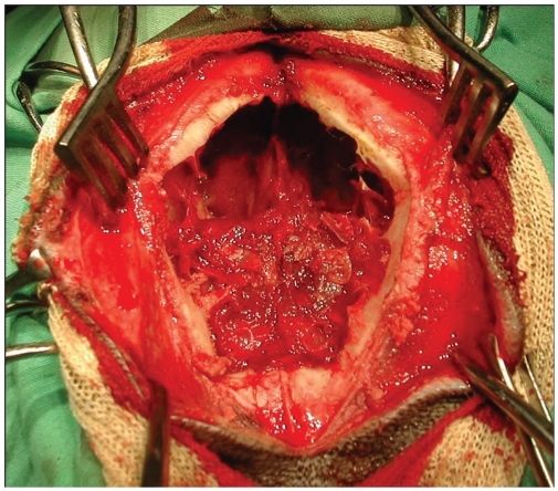
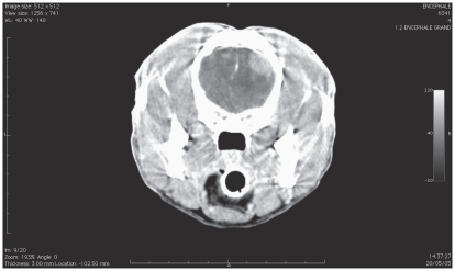
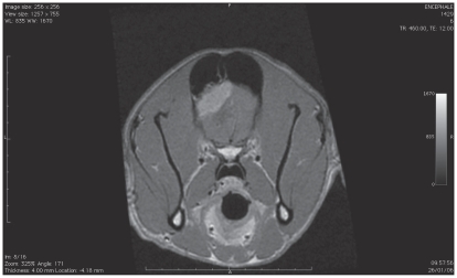
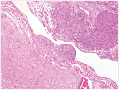
The cases of 7 adult dogs with generalized seizures managed by surgical excision and radiation therapy for frontal lobe meningiomas were reviewed. The neurological examination was unremarkable in 6 of the 7 dogs. Five dogs were operated on using a bilateral transfrontal sinus approach and 2 using a unilateral sinotemporal approach to the frontal lobe. One dog was euthanized 14 d after surgery; radiation therapy was initiated 3 wk after surgery in the remaining 6 dogs. Long-term follow-up consisted of neurological examination and magnetic resonance imaging (MRI) and/or computed tomography (CT) scan after radiation therapy. The mean survival time for dogs that had surgery and radiation therapy was 18 mo after surgery. Frontal lobe meningiomas have been associated with poor prognosis. However, the surgical approaches used in these cases, combined with radiation therapy, allow a survival rate for frontal lobe meningiomas similar to that for meningiomas located over the cerebral convexities.
Traitement chirurgical et radiothérapie de méningiomes du lobe frontal chez 7 chiens. Les cas de 7 chiens adultes avec des crises d’épilepsie généralisées gérées par ablation chirurgicale et radiothérapie pour des méningiomes du lobe frontal ont été examinés. L’examen neurologique était normal chez 6 des 7 chiens. Cinq chiens ont été opérés à l’aide d’une approche par le sinus transfrontal bilatéral et 2 à l’aide d’une approche sinotemporale unilatérale du lobe frontal. Un chien a été euthanasié 14 jours après la chirurgie; la radiothérapie a été entamée 3 semaines après la chirurgie chez les autres 6 chiens. Le suivi à long terme se composait d’un examen neurologique et d’une IRM et/ou d’un tomodensitogramme après la radiothérapie. La durée moyenne de survie pour les chiens qui avaient subi la chirurgie et la radiothérapie était de 18 mois après la chirurgie. Les méningiomes du lobe frontal ont été associés à un pronostic sombre. Cette étude documente que les approches chirurgicales utilisées pour ces cas, combinées à une radiothérapie, permettent un taux de survie pour les méningiomes du lobe frontal semblable à celui des méningiomes situés sur les convexités cérébrales.
(Traduit par Isabelle Vallières)
Figures
Similar articles
-
A modified bilateral transfrontal sinus approach to the canine frontal lobe and olfactory bulb: surgical technique and five cases.J Am Anim Hosp Assoc. 2000 Jan-Feb;36(1):43-50. doi: 10.5326/15473317-36-1-43. J Am Anim Hosp Assoc. 2000. PMID: 10667405
-
Surgery alone or in combination with radiation therapy for treatment of intracranial meningiomas in dogs: 31 cases (1989-2002).J Am Vet Med Assoc. 2002 Dec 1;221(11):1597-600. doi: 10.2460/javma.2002.221.1597. J Am Vet Med Assoc. 2002. PMID: 12479332
-
A surgical approach to the canine olfactory bulb for meningioma removal.Vet Surg. 1987 Jul-Aug;16(4):273-7. doi: 10.1111/j.1532-950x.1987.tb00952.x. Vet Surg. 1987. PMID: 3507155
-
External beam radiation therapy for canine intracranial meningioma.Can Vet J. 2009 Jan;50(1):97-100. Can Vet J. 2009. PMID: 19337624 Free PMC article. Review. No abstract available.
-
Intracranial meningioma in dogs and cats: a comparative review.Compend Contin Educ Vet. 2009 Jul;31(7):330-9. Compend Contin Educ Vet. 2009. PMID: 19746352 Review.
Cited by
-
Formation of a meningoencephalocele after removal of a frontal lobe meningioma by transfrontal craniotomy in a cat.JFMS Open Rep. 2020 Sep 15;6(2):2055116920957195. doi: 10.1177/2055116920957195. eCollection 2020 Jul-Dec. JFMS Open Rep. 2020. PMID: 32983552 Free PMC article.
-
Endoscopy-assisted resection of a sphenoid-wing meningioma using a 3D-printed patient-specific pointer in a dog: A case report.Front Vet Sci. 2022 Nov 16;9:979290. doi: 10.3389/fvets.2022.979290. eCollection 2022. Front Vet Sci. 2022. PMID: 36467647 Free PMC article.
-
Therapeutic effect of nerve growth factor on cerebral infarction in dogs using the hemisphere anomalous volume ratio of diffusion-weighted magnetic resonance imaging.Neural Regen Res. 2012 Aug 25;7(24):1873-80. doi: 10.3969/j.issn.1673-5374.2012.24.005. Neural Regen Res. 2012. PMID: 25624813 Free PMC article.
-
Comparison of Canine and Feline Meningiomas Using the Apparent Diffusion Coefficient and Fractional Anisotropy.Front Vet Sci. 2021 Jan 11;7:614026. doi: 10.3389/fvets.2020.614026. eCollection 2020. Front Vet Sci. 2021. PMID: 33506001 Free PMC article.
-
Volumetric Modulated Arc (Radio) Therapy in Pets Treatment: The "La Cittadina Fondazione" Experience.Cancers (Basel). 2018 Jan 24;10(2):30. doi: 10.3390/cancers10020030. Cancers (Basel). 2018. PMID: 29364837 Free PMC article. Review.
References
-
- Heidner GL, Kornegay JN, Page RL, et al. Analysis of survival in a retrospective study of 86 dogs with brain tumors. J Vet Int Med. 1991;5:219–26. - PubMed
-
- Snyder JM, Shofer FS, Winkle JV, et al. Canine intracranial primary neoplasia: 173 cases (1986–2003) J Vet Intern Med. 2006;20:669–675. - PubMed
-
- Adamo PF, Forrest L, Dubielzig R. Canine and feline meningiomas: Diagnosis, treatment and prognosis. Compend Contin Educ Pract Vet. 2004;26:951–964.
-
- Foster ES, Carrillo JM, Patnaik AK. Clinical signs of tumors affecting the rostral cerebrum in 43 dogs. J Vet Int Med. 1988;2:71–74. - PubMed
-
- Patnaik AK, Lieberman PH, Erlandson RA, et al. Paranasal meningioma in the dog: A clinicopathologic study of ten cases. Vet Path. 1986;23:369–373. - PubMed
MeSH terms
LinkOut - more resources
Full Text Sources