. 2012 Feb 20;25(2):454-61.
doi: 10.1021/tx200494h. Epub 2012 Jan 24.

Selection of monoclonal antibodies against 6-oxo-M(1)dG and their use in an LC-MS/MS assay for the presence of 6-oxo-M(1)dG in vivo

Affiliations

Selection of monoclonal antibodies against 6-oxo-M(1)dG and their use in an LC-MS/MS assay for the presence of 6-oxo-M(1)dG in vivo

Dapo Akingbade et al. Chem Res Toxicol. .

Abstract

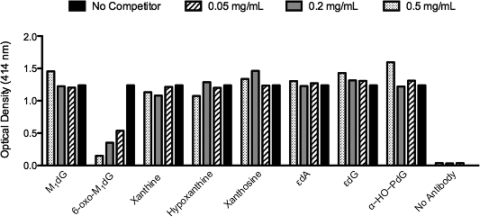
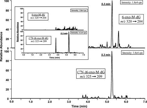
Oxidative stress triggers DNA and lipid peroxidation, leading to the formation of electrophiles that react with DNA to form adducts. A product of this pathway, (3-(2'-deoxy-β-d-erythro-pentofuranosyl)-pyrimido[1,2-α]purine-10(3H)-one), or M(1)dG, is mutagenic in bacterial and mammalian cells and is repaired by the nucleotide excision repair pathway. In vivo, M(1)dG is oxidized to a primary metabolite, (3-(2-deoxy-β-d-erythro-pentofuranosyl)-pyrimido[1,2-α]purine-6,10(3H,5H)-dione, or 6-oxo-M(1)dG, which is excreted in urine, bile, and feces. We have developed a specific monoclonal antibody against 6-oxo-M(1)dG and have incorporated this antibody into a procedure for the immunoaffinity isolation of 6-oxo-M(1)dG from biological matrices. The purified analyte is quantified by LC-MS/MS using a stable isotope-labeled analogue ([(15)N(5)]-6-oxo-M(1)dG) as an internal standard. Healthy male Sprague-Dawley rats excreted 6-oxo-M(1)dG at a rate of 350-1893 fmol/kg·d in feces. This is the first report of the presence of the major metabolite of M(1)dG in rodents without exogenous introduction of M(1)dG.


Figures

Scheme 1
Scheme 1. Conjugation Reaction between 6-oxo-M1Guo and the Carrier Protein (BSA or mcKLH)
Figure 1
Figure 1
6-oxo-M1dG, M1dG, and structural analogues that were used as competitive antigens during ELISA analysis of murine sera, hybridomal supernatant, and purified antibodies.
Figure 2
Figure 2
Schematic of monoclonal antibody generation and isolation procedure. Eight mice were inoculated with adduct/protein conjugate, followed by tail bleeding and testing for the presence of 6-oxo-M1dG antibodies using direct ELISA analysis (step 1). Splenocytes from a single mouse, Balb/CJ R, were fused with myeloma cells, plated on twenty-four 96-well plates, and screened using direct and competitive ELISA analyses (step 2). From this screen, 180 hybridoma cell lines showed anti-6-oxo-M1dG mAb production. These were replated and screened (step 3). Five parental cell lines were chosen for subcloning and further screening (step 4). On the basis of these final screenings, the cell line 6C9BA4C12 was chosen for scale-up and antibody purification (step 5).
Figure 3
Figure 3
6-oxo-M1dG antibodies are present in the serum of mouse BALB/cJ R. The serum of mouse BALB/cJ R was subjected to competitive ELISA analysis with 6-oxo-M1dG used as the competitor. The displayed results represent the average of spectroscopic readings at 15 and 30 min postaddition of ABTS substrate.
Figure 4
Figure 4
ELISA analysis of specificity of various subclonal antibodies. The specificity of antibodies produced by two hybridoma cell lines (B and E2) subcloned from the parental hybridoma cell line 6C9 were assessed using competitive ELISA analysis. Xanthine and 6-oxo-M1dG were used as competing antigens. The displayed results represent the average of spectroscopic readings of subclones E2 and B at 15 and 30 min postaddition of ABTS substrate.
Figure 5
Figure 5
Specificity determination of antibodies produced from subclone 6C9BA4C12. Purified antibodies from the hybridomal subclone (6C9BA4C12) were screened for specificity for 6-oxo-M1dG in the presence of structural analogues using competitive ELISA analyses.
Figure 6
Figure 6
6-oxo-M1dG is present in rat feces. A representative LC-MS/MS chromatogram of 6-oxo-M1dG and the internal standard [15N5]-6-oxo-M1dG isolated from rat feces is displayed. The inset represents a chromatogram of internal standard alone, purified from PBS.

References

    1. Basu A. K.; O’Hara S. M.; Valladier P.; Stone K.; Mols O.; Marnett L. J. (1988) Identification of adducts formed by reaction of guanine nucleosides with malondialdehyde and structurally related aldehydes. Chem. Res. Toxicol. 1, 53–59. - PubMed
    1. Chaudhary A. K.; Nokubo M.; Reddy G. R.; Yeola S. N.; Morrow J. D.; Blair I. A.; Marnett L. J. (1994) Detection of endogenous malondialdehyde-deoxyguanosine adducts in human liver. Science 265, 1580–1582. - PubMed
    1. Rouzer C. A.; Chaudhary A. K.; Nokubo M.; Ferguson D. M.; Reddy G. R.; Blair I. A.; Marnett L. J. (1997) Analysis of the malondialdehyde-2′-deoxyguanosine adduct pyrimidopurinone in human leukocyte DNA by gas chromatography/electron capture/negative chemical ionization/mass spectrometry. Chem. Res. Toxicol. 10, 181–188. - PubMed
    1. Fink S. P.; Reddy G. R.; Marnett L. J. (1997) Mutagenicity in Escherichia coli of the major DNA adduct derived from the endogenous mutagen malondialdehyde. Proc. Natl. Acad. Sci. U.S.A. 94, 8652–8657. - PMC - PubMed
    1. VanderVeen L. A.; Hashim M. F.; Shyr Y.; Marnett L. J. (2003) Induction of frameshift and base pair substitution mutations by the major DNA adduct of the endogenous carcinogen malondialdehyde. Proc. Natl. Acad. Sci. U.S.A. 100, 14247–14252. - PMC - PubMed

Publication types