. 2012 May;22(5):859-72.
doi: 10.1038/cr.2012.3. Epub 2012 Jan 3.

A distinct response to endogenous DNA damage in the development of Nbs1-deficient cortical neurons

Affiliations

A distinct response to endogenous DNA damage in the development of Nbs1-deficient cortical neurons

Rui Li et al. Cell Res. 2012 May.

Abstract

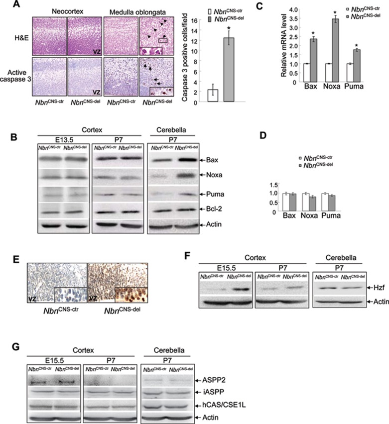
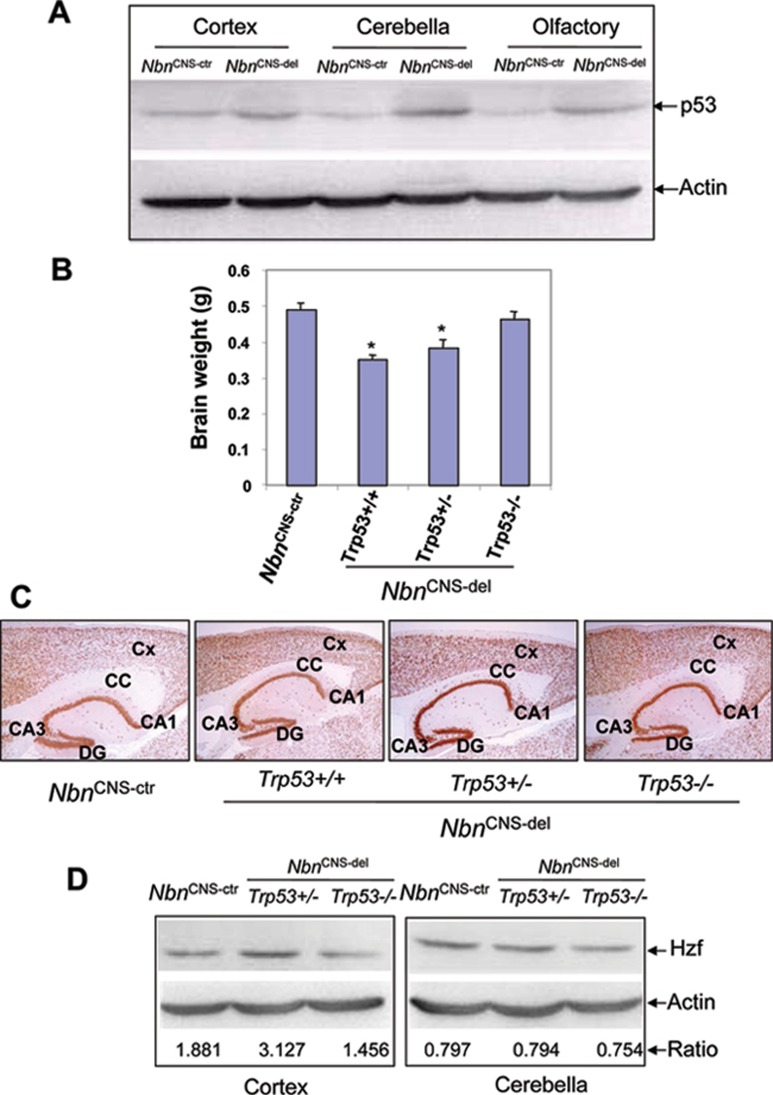
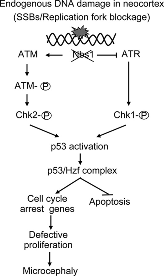
Microcephaly is a clinical characteristic for human nijmegen breakage syndrome (NBS, mutated in NBS1 gene), a chromosomal instability syndrome. However, the underlying molecular pathogenesis remains elusive. In the present study, we demonstrate that neuronal disruption of NBS (Nbn in mice) causes microcephaly characterized by the reduction of cerebral cortex and corpus callosum, recapitulating neuronal anomalies in human NBS. Nbs1-deficient neocortex shows accumulative endogenous DNA damage and defective activation of Ataxia telangiectasia and Rad3-related (ATR)-Chk1 pathway upon DNA damage. Notably, in contrast to massive apoptotic cell death in Nbs1-deficient cerebella, activation of p53 leads to a defective neuroprogenitor proliferation in neocortex, likely via specific persistent induction of hematopoietic zinc finger (Hzf) that preferentially promotes p53-mediated cell cycle arrest whilst inhibiting apoptosis. Moreover, Trp53 mutations substantially rescue the microcephaly in Nbs1-deficient mice. Thus, the present results reveal the first clue that developing neurons at different regions of brain selectively respond to endogenous DNA damage, and underscore an important role for Nbs1 in neurogenesis.


Figures

Figure 1
Figure 1
Nbs1 deficiency leads to microcephaly. (A) Macroscopic view of reduced cerebral size in NbnCNS-del brain, n = 20, P < 0.01 (t test). (B) NeuN staining of coronal section of the cerebrum at postnatal day 21 (P21), original magnification ×1. Cx, cerebral cortex; CC, corpus callosum; H, hippocampus; fc, fasciole cinerum; Mhb, medial habenular neurons; T, thalamic region; HT, hypothalamic region. (C) Representative laminar cortical structure in control and NbnCNS-del mice. (D) Histogram showing the percentage of neurons in each layer of NbnCNS-del cerebral cortical sections compared to control mice. At least 10 mice of each genotype were analyzed, *P < 0.01 (t test). (E) Representative P7 hippocampus with NeuN staining (left). DG, dentate gyrus; CA1/CA3, field CA1/CA3 of hippocampus; PoDG, polymorph layer dentate gyrus. Bar is 1 mm. Representative MBP staining for CC and neurofibers (right). (F) Quantification of the thickness of CC in NbnCNS-del brain at the indicated site. Original magnification ×10. At least 10 mice of each genotype were analyzed, *P < 0.01 (t test).
Figure 2
Figure 2
Nbs1 deficiency impairs ATR-Chk1 signaling in response to endogenous DNA damage in developing brain. (A) Accumulation of γH2AX exists in VZ and MZ of Nbs1-deficient embryonic brain at E12.5. Original magnification ×40. (B) Schematic of the role of Nbs1 in response to DNA damage. (C) Nbs1 is required for the activation of ATR signaling during neurogenesis. Western blot analysis of the activation of ATR pathway in embryonic cortex. Note: In the absence of Nbs1, a marked reduction of Chk1-S345p at E15.5–18.5 during neurogenesis.
Figure 3
Figure 3
Nbs1 deficiency impairs phosphorylated Chk1 focus formation. Primary cultured NbnCNS-ctr and NbnCNS-del developing neurons were treated with 2 mM HU for 3 h, and subjected to immunoflurescence staining with antibodies against phosphorylated Chk1-S345p (A), BRCA1 (B), γH2AX (C), and Nbs1 partner Mre11 (D). DAPI stains nuclei. The neuroprogenitors (A-D) are verified by nestin-positive immunoactivity, and at least 150 cells per staining were analyzed.
Figure 4
Figure 4
Nbs1 deficiency leads to defects in proliferation of neuroprogenitors during neurogenesis. (A) Activation of the p53 pathway in Nbs1-deficient embryonic cortex. Representative immunostaining of p53 and its downstream target p21 at E12.5. Original magnification ×40. Insets: positive p53 and p21 cells. (B) Western blot analysis of the activation of p53 protein in E15.5 neocortex. (C) Nbs1 deficiency results in increased expression level of p53 target gene p21 in E15.5 neocortex and in P7 cerebella (as a positive control of inhibition of cell proliferation). (D) Representative in vivo BrdU labeling at E12.5. (E) Histogram representing percentage of BrdU-positive cells at VZ of 6 high-power fields from at least five mice of each genotype at E12.5. *P < 0.01 (t test).
Figure 5
Figure 5
Role of Nbs1 in neuronal cell death. (A) H&E and active caspase 3 immunostaining showing cell death in medulla oblongata at E12.5, but not in developing cortex at E15.5. Histogram representing percentage of active caspase 3-positive cells at medulla oblongata of six high-power fields from at least eight mice of each genotype at E12.5 (Original magnification ×40). *P < 0.001 (t test). (B) Nbs1 deficiency does not affect the expression of p53-dependent proapoptotic protein Bax, Noxa and Puma in neocortex at E13.5 and cerebral cortex at P7, but loss of Nbs1 alters cerebellar proapoptotic protein Bax, Noxa and Puma at P7. (C) Nbs1 deficiency results in increased expression levels of p53-mediated proapoptotic target genes Bax, Noxa and Puma in P7 cerebella, but has no effect on these gene expression in neocortex at E15.5 (D). At least six mice of each genotype were analyzed, *P < 0.05 (t test). (E) Hzf immunoactivity in Nbs1-proficient and -deficient neocortex at E15.5 and E12.5 (inset). Original magnification ×40. Insets: positive Hzf cells in VZ region. (F) Expression level of Hzf in E15.5, P7 Nbs1-proficient and -deficient cortex, and P7 cerebella. (G) Expression of ASPP2, iASPP and hCAS/CSE1L proteins in E15.5, P7 Nbs1-proficient and -deficient cortex, and P7 cerebella.
Figure 6
Figure 6
Introduction of Trp53 mutations in Nbs1-deficient brain largely rescues microcephaly. (A) Western blot analysis of p53 protein in Nbs1-deficient and -proficient brains at P7. (B) Brain weight from at least 10 mice of each genotype. *P < 0.01 (t test). (C) Histological analysis of Trp53 mutant Nbs1-deficient cerebrum. Cx, cerebral cortex; CC, corpus callosum; DG, dentate gyrus; CA1/CA3, field CA1/CA3 of hippocampus. Original magnification ×4. (D) Expression level of Hzf protein in P7 Trp53 mutant Nbs1-deficient cortex and cerebella.
Figure 7
Figure 7
Working model for Nbs1 function and regulation in neocortex. Endogenous DNA damage during rapid development of neocortical neurons activates ATM-Chk2 and ATR-Chk1 signaling. However, defective Nbs1 impairs ATR-Chk1 pathway rather than ATM-Chk2 signaling. ATM-Chk2 signaling in Nbs1-deficient neocortex activates p53-Hzf pathway and induces defective proliferation rather than apoptosis of neuroprogenitors, leading to microcephaly.

References

    1. Varon R, Vissinga C, Platzer M, et al. Nibrin, a novel DNA double-strand break repair protein, is mutated in Nijmegen breakage syndrome. Cell. 1998;93:467–476. - PubMed
    1. Riches LC, Lynch AM, Gooderham NJ. Early events in the mammalian response to DNA double-strand breaks. Mutagenesis. 2008;23:331–339. - PubMed
    1. Lee JH, Paull TT. ATM activation by DNA double-strand breaks through the Mre11-Rad50-Nbs1 complex. Science. 2005;308:551–554. - PubMed
    1. Difilippantonio S, Nussenzweig A. The NBS1-ATM connection revisited. Cell Cycle. 2007;6:2366–2370. - PubMed
    1. Jazayeri A, Balestrini A, Garner E, Haber JE, Costanzo V. Mre11-Rad50-Nbs1-dependent processing of DNA breaks generates oligonucleotides that stimulate ATM activity. EMBO J. 2008;27:1953–1962. - PMC - PubMed

Publication types

MeSH terms