. 2012 Sep;16(9):2140-9.
doi: 10.1111/j.1582-4934.2012.01524.x.

Identification of F-box only protein 7 as a negative regulator of NF-kappaB signalling

Affiliations

Identification of F-box only protein 7 as a negative regulator of NF-kappaB signalling

Hendrik J Kuiken et al. J Cell Mol Med. 2012 Sep.

Abstract

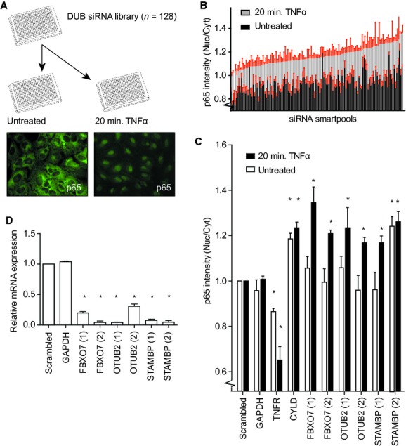
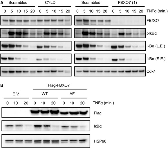
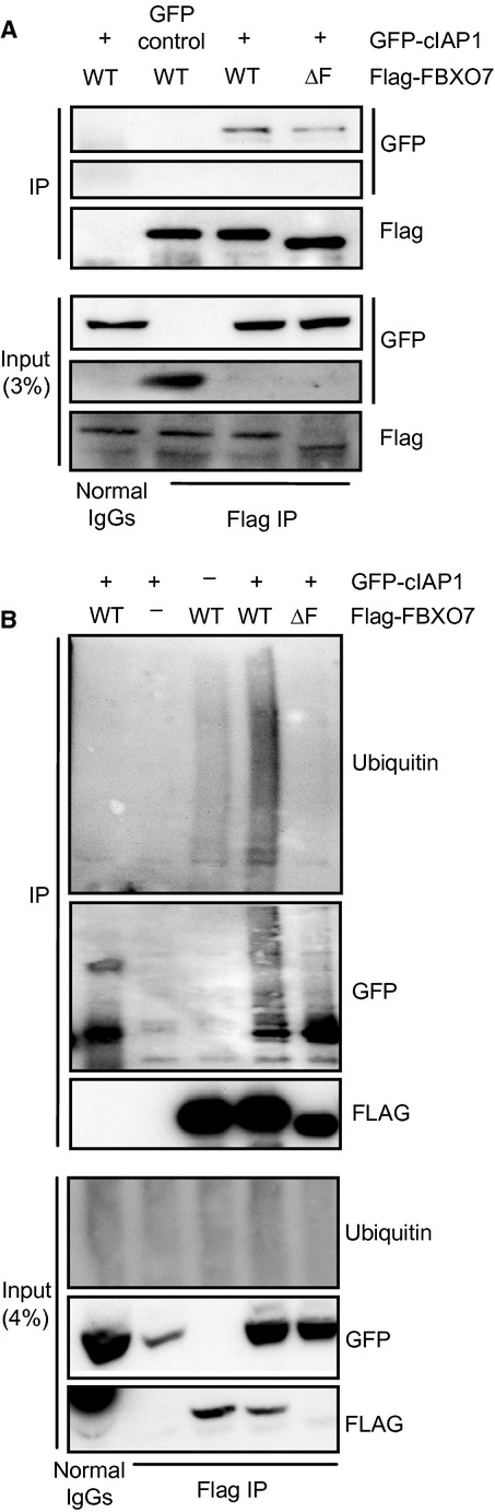
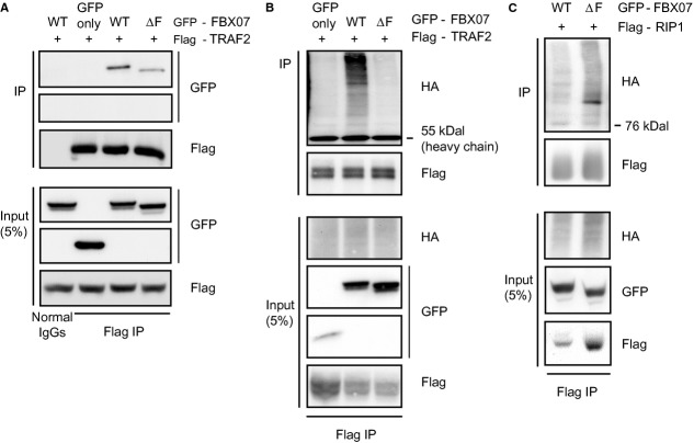
The nuclear factor κB (NF-κB) signalling pathway controls important cellular events such as cell proliferation, differentiation, apoptosis and immune responses. Pathway activation occurs rapidly upon TNFα stimulation and is highly dependent on ubiquitination events. Using cytoplasmic to nuclear translocation of the NF-κB transcription factor family member p65 as a read-out, we screened a synthetic siRNA library targeting enzymes involved in ubiquitin conjugation and de-conjugation for modifiers of regulatory ubiquitination events in NF-κB signalling. We identified F-box protein only 7 (FBXO7), a component of Skp, Cullin, F-box (SCF)-ubiquitin ligase complexes, as a negative regulator of NF-κB signalling. F-box protein only 7 binds to, and mediates ubiquitin conjugation to cIAP1 and TRAF2, resulting in decreased RIP1 ubiquitination and lowered NF-κB signalling activity.


Figures

Fig 1
Fig 1
Screen for modulators of NF-κB signalling. (A) Screen setup: U2OS cells were transfected in a single well format with the DUB siRNA library (Thermo Scientific), stimulated with TNFα or left untreated, and analysed for cellular redistribution of NF-κB p65. Representative photos of unstimulated and TNFα-stimulated cells showing endogenous p65 staining. (B) Control normalized ratios of nuclear over cytoplasmic p65 intensity for the siRNA smartpools included in the screen. Error bars shown in red. (C) Summary of three independent experiments for cellular redistribution of p65 upon knockdown of FBXO7, OTUB2, STAMBP, and control genes, GAPDH, TNF-R1 and CYLD. A non-targeting (scrambled) siRNA smartpool was used as negative control. (D) Summary of three independent experiments showing the knockdown efficiency determined by qRT PCR for mRNA levels of the identified genes. (C and D) *P < 0.05, values are mean ± standard deviation. P-value computed from unpaired two-tailed t-test.
Fig 2
Fig 2
FBXO7 regulates NF-κB signalling. (A and B) NF-κB target gene, A20 and IL8, activation determined by qRT PCR upon stimulation with TNFα in (A) U2OS and (B) immortalized primary human fibroblasts (BJET) transfected with siRNAs targeting FBXO7, TNF-R1, CYLD or a non-targeting (scrambled) control. Values are mean ± standard deviation of replicate measurements from a representative experiment. (C) NF-κB luciferase reporter assay in U2OS cells expressing the NF-κB luciferase reporter construct, a SV40-Renilla construct, and either empty vector (pcDNA3.1), Flag-tagged wild-type FBXO7 (WT), or Flag-tagged F-box deletion mutant FBXO7 (ΔF). Values represent ratio of Luciferase activity over Renilla control activity in the absence or presence of TNFα. *P < 0.05, values are mean ± standard deviation of four independent experiments. P-value computed from unpaired two-tailed t-test.
Fig 3
Fig 3
FBXO7 regulates the NF-κB signalling upstream of IκBα degradation. (A) Western blot analysis of U2OS cells transfected with non-targeting (scrambled) control, CYLD or FBXO7 siRNAs stimulated with TNFα for 5, 10, 15, 20 min., or left untreated. Western blot analysis was performed for FBXO7, pIκBα, IκBα and Cdk4 (loading control). L.E.: long exposure; S.E.: short exposure. (B) Western blot analysis of U2OS cells expressing empty vector (EV), Flag-FBXO7 WT, or ΔF. Western blot analysis was performed for the Flag epitope, IκBα and HSP90 (loading control).
Fig 4
Fig 4
FBXO7 interacts with and mediates ubiquitination of cIAP1. (A) Western blot analysis of a co-immunoprecipitation experiment in U2OS cells expressing GFP-cIAP1 and Flag-FBXO7 WT or ΔF. (B) Western blot analysis of in vivo ubiquitinated GFP-cIAP1 co-immunoprecipitated with Flag-FBXO7 WT or ΔF.
Fig 5
Fig 5
FBXO7 interacts with TRAF2, and alters ubiquitination of TRAF2 and RIP1. (A) Western blot analysis of a co-immunoprecipitation experiment in U2OS cells expressing Flag-TRAF2 and GFP-tagged wild-type FBXO7 WT or ΔF. (B and C) Western blot analysis of an in vivo ubiquitination assays in HEK293 cells expressing HA-ubiquitin, Flag-TRAF2 or Flag-RIP1, GFP only, GFP-FBXO7 WT or ΔF.

References

    1. Chen G, Goeddel DV. TNF-R1 signaling: a beautiful pathway. Science. 2002;296:1634–5. - PubMed
    1. Karin M, Cao Y, Greten FR, et al. NF-kappaB in cancer: from innocent bystander to major culprit. Nat Rev Cancer. 2002;2:301–10. - PubMed
    1. Chan FK. Three is better than one: pre-ligand receptor assembly in the regulation of TNF receptor signaling. Cytokine. 2007;37:101–7. - PMC - PubMed
    1. Wajant H, Scheurich P. TNFR1-induced activation of the classical NF-kappaB pathway. FEBS J. 2011;278:862–76. - PubMed
    1. Vince JE, Pantaki D, Feltham R, et al. TRAF2 must bind to cellular inhibitors of apoptosis for tumour necrosis factor (tnf) to efficiently activate nf-{kappa}b and to prevent tnf-induced apoptosis. J Biol Chem. 2009;284:35906–15. - PMC - PubMed

Publication types

MeSH terms