. 2012 Aug 1;72(11):1214-22.
doi: 10.1002/pros.22472. Epub 2011 Dec 27.

Deficiency of DNA repair nuclease ERCC1-XPF promotes prostate cancer progression in a tissue recombination model

Affiliations

Deficiency of DNA repair nuclease ERCC1-XPF promotes prostate cancer progression in a tissue recombination model

Derek J Matoka et al. Prostate. .

Abstract

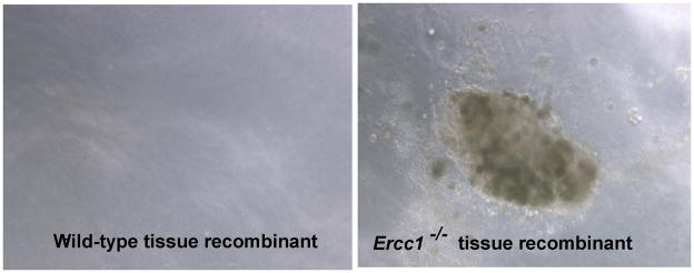
Background: The excision repair cross complementing (ERCC1) gene product plays a vital role in the nucleotide excision repair (NER) and DNA interstrand crosslink repair pathways, which protect the genome from mutations and chromosomal aberrations, respectively. Genetic deletion of Ercc1 in the mouse causes dramatically accelerated aging. We examined the effect of Ercc1 deletion in the development of prostate cancer in a prostate recapitulation model as Ercc1 deficient mice die within four weeks of birth.

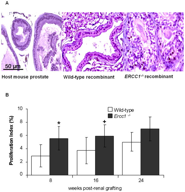
Methods: Prostate tissues from Ercc1(-/-) mice or wild-type littermates were combined with embryonic rat urogenital mesenchyme and grown as renal grafts for a total of 8, 16, and 24 weeks before histological, expression and proliferative evaluation.

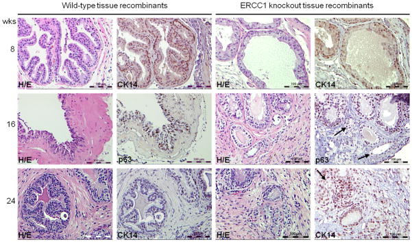
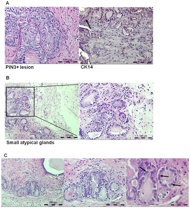
Results: Invasive adenocarcinoma was observed in Ercc1(-/-) tissue recombinants but not wild-type as early as 8 weeks post-grafting. PIN-like lesions in Ercc1(-/-) tissue recombinants had more cytologic and architectural atypia than wild-type (P = 0.02, P = 0.0065, and P = 0.0003 at the 8, 16, and 24 weeks, respectively), as well as more proliferative cells (P = 0.022 and P = 0.033 at 8 and 16 weeks, respectively). With serial grafting, Ercc1(-/-) tissue recombinants progressed to a more severe histopathological phenotype more rapidly than wild-type (P = 0.011).

Conclusions: Results show that ERCC1 and by implication the NER and/or interstrand crosslink repair mechanisms protect against prostate carcinogenesis and mutations or polymorphisms affecting these DNA repair pathways may predispose prostate epithelial cells to transformation.


Conflict of interest statement

Disclosure Statement: The authors declare that they have no affiliations with any organization that may have a direct interest in the research described, or a real or perceived conflict of interest. The funders had no role in study design, data collection and analysis, decision to publish, or preparation of the manuscript.

Figures

Figure 1
Figure 1
Histological comparison of wild-type and Ercc1−/− tissue recombinants treated with T&E2 at 8 weeks post-renal grafting. (A) Prostate tissue from host Nude mouse. (B) Wild-type tissue recombinants displayed normal glandular architecture and focal hyperplasia. Ercc1−/− tissue recombinants displayed (C) 2+ PIN-like lesions, and (D) focal areas of nuclear pleomorphism, abnormal mitosis and crowding with epithelial proliferation. Arrows point to cells in mitoses. (A–D, scale bar = 50 μm) (E) Tissue sections were stained with Hoechst 33258 stain, with the nucleus of mouse origin having a spotty appearance while the cells of rat origin have a diffuse appearance. (left, scale bar = 100 μm; right, scale bar = 33 μm)
Figure 2
Figure 2
Comparison of tissue recombinants at 8, 16 and 24 weeks post initial renal grafting. Small atypical glands with features of adenocarcinoma were observed only in Ercc1−/− tissue recombinants. H/E and CK-14/p63 staining of wild-type and Ercc1−/− tissue recombinants at 8, 16 and 24 weeks after renal grafting in immunodeficient hosts treated with T&E2. Arrows point to small atypical glands that lack of CK-14/p63-staining, indicative of adenocarcinoma (magnification ×20, scale bar = 100 μm).
Figure 3
Figure 3
Proliferation index of Ercc1−/− and wild-type tissue recombinants at 8 and 16 weeks post-renal grafting. (A) Histology of Ki67 stained host mouse prostate; wild-type, and Ercc1−/− prostate tissue recombinants at 16 weeks post-renal grafting (magnification ×20, scale bar = 50 μm). (B) Proliferation index of wild-type and Ercc1−/− tissue recombinants at 8, 16 and 24 post-renal grafting. Proliferation was significantly higher in Ercc1−/− tissue recombinants at 8 (*p=0.022) and 16 (+p=0.033) weeks post-renal grafting. Data are presented as mean ± SEM.
Figure 4
Figure 4
Histology of Ercc1−/− tissue recombinants treated with T&E2 at 16 (A, B) and 24 (C) weeks post-renal grafting. (A) Ercc1−/− tissue recombinants displayed 3+ PIN-like lesion with hyperplasia, nuclear stratification, elevated nuclear to cytoplasmic ratios and papillary tufting and wisps of CK-14 positive (arrow) basal cells (scale bar = 100 μm; left and right panels are independent images). (B) Representative images of the same Ercc1−/− tissue recombinant with multiple, small, crowded glands with nuclear pleomorphism, elevated nuclear to cytoplasmic ratio, and abnormal mitosis – all characteristics indicative of carcinoma. (left, scale bar = 200 μm; right, scale bar = 100 μm) (C) Representative images of the same Ercc1−/− tissue recombinant at 24 weeks that displayed multiple small, crowded glands with large prominent nucleoli (arrows), high nuclear to cytoplasmic ratio, and abnormal mitosis (left, scale bar = 200 μm; middle, scale bar = 100 μm; right, scale bar = 50 μm).
Figure 5
Figure 5
Growth of cells from 24 week tissue recombinants in semisolid media. Cells isolated from wild-type tissue recombinants did not form colonies on semisolid medium. In contrast, cells isolated from Ercc1−/− tissue recombinants that had been grafted under the renal capsule for 24 weeks were able to form colonies on semisolid medium.

Similar articles

Cited by

References

    1. Jemal A, Siegel R, Xu J, Ward E. Cancer Statistics, 2010. CA Cancer J Clin. 2010;60:277–300. - PubMed
    1. Malins DC, Johnson PM, Wheeler TM, Barker EA, Polissar NL, Vinson MA. Age-related radical-induced DNA damage is linked to prostate cancer. Cancer Res. 2001;61:6025–6028. - PubMed
    1. Isaacs WB, Bova GS, Morton RA, Bussemakers MJ, Brooks JD, Ewing CM. Molecular biology of prostate cancer. Semin Oncol. 1994;21:514–521. - PubMed
    1. Coughlin SS, Hall IJ. A review of genetic polymorphisms and prostate cancer risk. Ann Epidemiol. 2002;12:182–196. - PubMed
    1. Dong JT. Prevalent mutations in prostate cancer. J Cell Biochem. 2006;97:433–447. - PubMed

Publication types