. 2012 Oct;227(10):3405-16.
doi: 10.1002/jcp.24041.

VE-cadherin trans-interactions modulate Rac activation and enhancement of lung endothelial barrier by iloprost

Affiliations

VE-cadherin trans-interactions modulate Rac activation and enhancement of lung endothelial barrier by iloprost

Anna A Birukova et al. J Cell Physiol. 2012 Oct.

Abstract

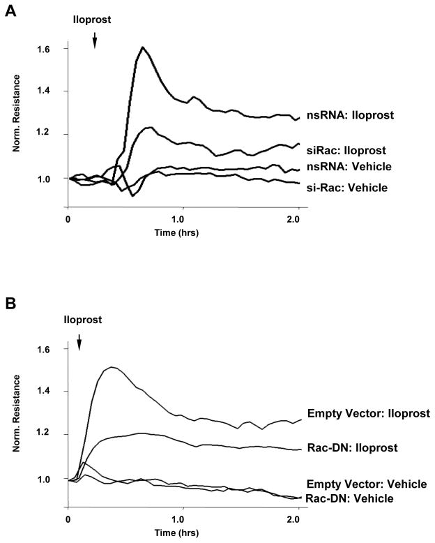
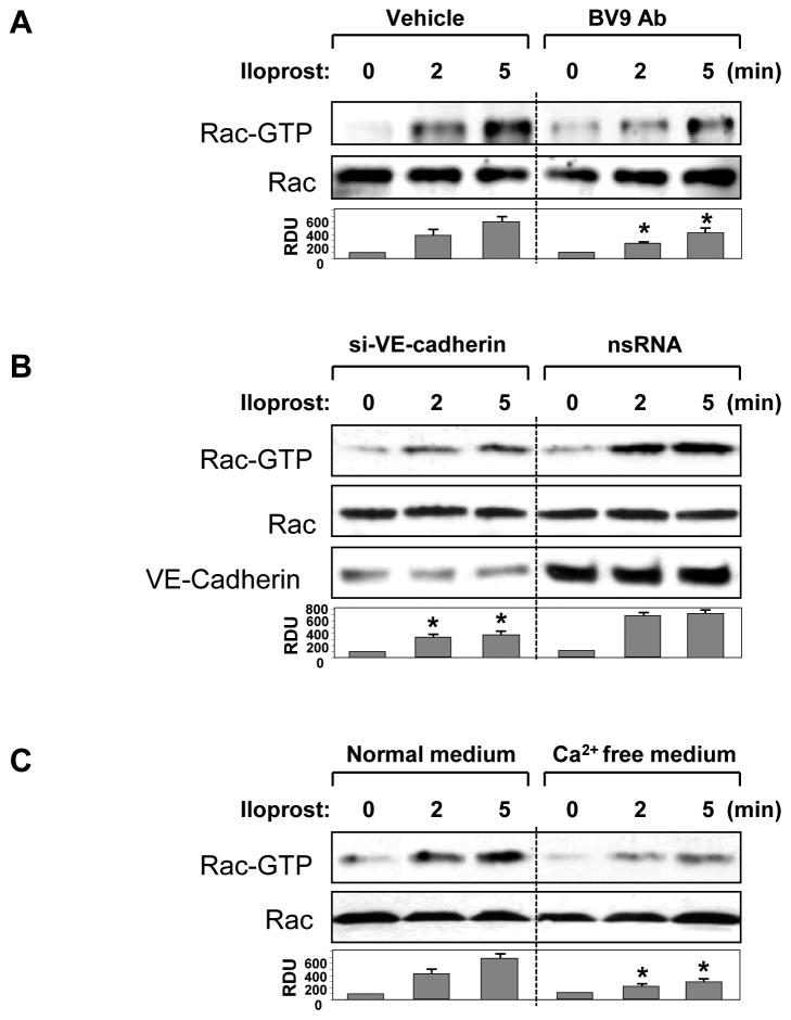
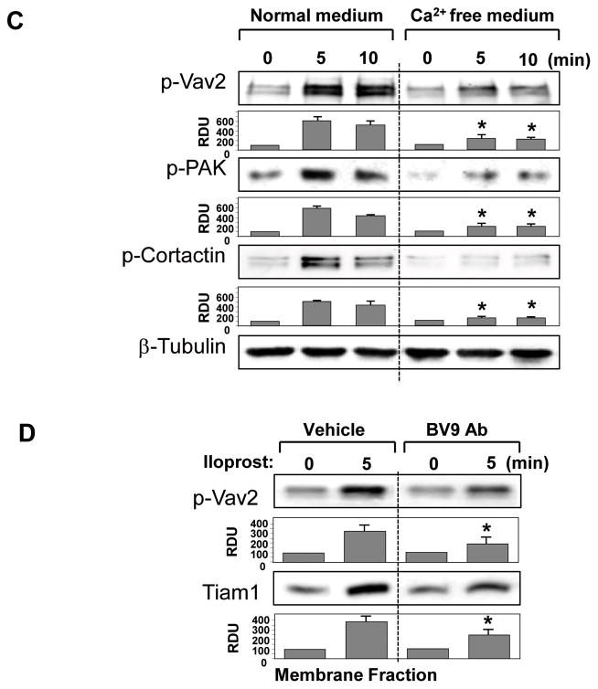
Small GTPase Rac is important regulator of endothelial cell (EC) barrier enhancement by prostacyclin characterized by increased peripheral actin cytoskeleton and increased interactions between VE-cadherin and other adherens junction (AJ) proteins. This study utilized complementary approaches including siRNA knockdown, culturing in Ca(2+) -free medium, and VE-cadherin blocking antibody to alter VE-cadherin extracellular interactions to investigate the role of VE-cadherin outside-in signaling in modulation of Rac activation and EC barrier regulation by prostacyclin analog iloprost. Spatial analysis of Rac activation in pulmonary EC by FRET revealed additional spike in iloprost-induced Rac activity at the sites of newly formed cell-cell junctions. In contrast, disruption of VE-cadherin extracellular trans-interactions suppressed iloprost-activated Rac signaling and attenuated EC barrier enhancement and cytoskeletal remodeling. These inhibitory effects were associated with decreased membrane accumulation and activation of Rac-specific guanine nucleotide exchange factors (GEFs) Tiam1 and Vav2. Conversely, plating of pulmonary EC on surfaces coated with extracellular VE-cadherin domain further promoted iloprost-induced Rac signaling. In the model of thrombin-induced EC barrier recovery, blocking of VE-cadherin trans-interactions attenuated activation of Rac pathway during recovery phase and delayed suppression of Rho signaling and restoration of EC barrier properties. These results suggest that VE-cadherin outside-in signaling controls locally Rac activity stimulated by barrier protective agonists. This control is essential for maximal EC barrier enhancement and accelerated barrier recovery.


Figures

Figure 1
Figure 1. Effect of iloprost on F-actin remodeling, and VE-cadherin distribution in human pulmonary EC
A and B: Endothelial cells grown on glass coverslips were stimulated with iloprost (75 nM, 10 min) followed by immunofluorescence staining with Texas Red phalloidin to detect actin filaments (A) or VE-cadherin antibody (B). C: Quantitative analysis of iloprost-induced VE-cadherin peripheral accumulation. Data are expressed as mean ± SD of five independent experiments; *p<0.05. D: HPAEC were incubated with vehicle, iloprost (75 nM), or thrombin (0.2 U/ml) for 60 min followed by measurements of permeability for FITC-labeled dextran. Permeability data are expressed as mean ± SD of four independent experiments; *p<0.05.
Figure 2
Figure 2. Effect of BV9 blocking antibody on iloprost-induced EC barrier enhancement
A: HPAEC plated on microelectrodes were treated with vehicle (IgG) or BV9 antibody (50 μg/ml) followed by TER measurements for 15 hr. Inset depicts TER values in the time window of BV9 pretreatment (30 min) used in this study. B: Left panel - HPAEC were pretreated with BV9 antibody followed by iloprost stimulation (75 nM). TER was monitored over 2 hrs. Right panel - TER increase caused by iloprost stimulation of EC pretreated with vehicle was taken as 100%. Pretreatment with irrelevant isotype-specific antibody (Cbl) was used as negative control. Pretreatment with BV9 significantly attenuated TER increase caused by iloprost stimulation. Data are expressed as mean ± SD of five independent experiments; *p<0.05.
Figure 3
Figure 3. Effect of BV9 blocking antibody on iloprost-induced cytoskeletal remodeling
A and B: Sub-confluent endothelial cells grown on glass coverslips were pretreated with BV9 antibody (50 μg/ml, 30 min) followed by iloprost stimulation (75 nM, 10 min). Immunofluorescence staining was performed with Texas Red phalloidin (A) or cortactin antibody (B). Insets represent high magnification images of cortactin peripheral accumulation.
Figure 4
Figure 4. Involvement of Rac in iloprost-induced EC barrier enhancement
A: EC were transfected with Rac1-specific siRNA or non-specific RNA. EC were stimulated with iloprost (75 nM) or vehicle at the time indicated by arrow, and TER changes were monitored over 2 hr. B: EC monolayers were subjected to transfection with dominant-negative Rac (Rac-DN). Cells transfected with empty vector served as controls. After 48 hrs of transfection, cells were stimulated with iloprost. Changes in endothelial permeability were monitored by measurements of TER.
Figure 5
Figure 5. Effect of inhibition VE-cadherin interactions on iloprost-induced Rac activation
A – C: HPAEC were pretreated with vehicle or BV9 (50 μg/ml, 30 min) (A), transfected with VE-cadherin-specific siRNA or non-specific RNA duplexes (B), or incubated in 2% FCS containing media with or without EGTA (5 mM, 30 min) (C). Next, EC were stimulated with iloprost (75 nM) for the indicated periods of time. Effect of iloprost on Rac activation was evaluated by Rac-GTP pulldown assay. Upper panel depicts the levels of activated, GTP-bound Rac, and the lower panel shows total Rac content in EC lysates. VE-cadherin depletion induced by specific siRNA duplexes was confirmed by western blot analysis of protein content in whole cell lysates. Result of densitometry shown as mean ± SD, * p<0.05 as compared to corresponding iloprost-stimulated controls.
Figure 6
Figure 6. FRET analysis of iloprost-induced spatial Rac activation
A – C: HPAEC were transfected with non-specific of VE-cadherin-specific siRNA duplexes for 24 hr followed by transfection with CFP/YPet-Rac biosensor for additional 24 hrs. FRET analysis was performed in iloprost-stimulated (75 nM) cells under control conditions (A), in HPAEC pretreated with BV9 antibody (50 μg/ml, 30 min) (B), or in VE-cadherin depleted EC (C). Images represent ratio of activated Rac to the total Rac content. Areas of Rac activation appear in yellow. Higher magnification inset in the lower frame of panel depicts increased local Rac activation at the cell junction of iloprost-stimulated cells. D: Quantitative analysis of iloprost-induced Rac activation at the areas of cell-cell contacts. Bar graphs represent normalized (cell-cell contacts/cell center) CFP/YPet emission ratio. Peripheral Rac activation in cells without iloprost stimulation was compared to Rac activation after 4 min of iloprost addition. In experiments with VE-cadherin knockdown or VE-cadherin blocking antibody Rac activation was compared to iloprost-stimulated controls (4 min). Data are expressed as mean ± SD of four independent experiments, 5–7 cells for each experiment; *p<0.05.
Figure 7
Figure 7. Effect of inhibition of VE-cadherin interactions on iloprost-induced activation of Rac signaling
A – C: HPAEC were pretreated with vehicle or BV9 (50 μg/ml, 30 min) (A); transfected with VE-cadherin-specific or non-specific RNA (B); or incubated in 2% FCS culture medium with or without EGTA (5 mM, 30 min) (C). Cells were next stimulated with iloprost (75 nM) for the indicated periods of time. Effect of iloprost on Vav2, PAK, and cortactin phosphorylation was evaluated by western blot with corresponding antibodies. VE-cadherin depletion induced by specific siRNA duplexes was confirmed by western blot analysis of protein content in whole cell lysates. Equal protein loading was confirmed by membrane probing with β-tubulin antibody. D: The membrane fraction was isolated from iloprost-treated EC with or without BV9 pretreatment. Content of phosphorylated Vav2 or Tiam1 was determined by western blot with specific antibodies. Result of densitometry shown as mean ± SD, * p<0.05 as compared to corresponding iloprost-stimulated controls.
Figure 8
Figure 8. Role of VE-cadherin trans-interactions in iloprost-induced Rac signaling assessed by adhesion assay
Plastic plates were coated with Fc-VE-cadherin or control Fc fragments as described in Methods. HPAEC were allowed to attach to the matrices during 30 min followed by stimulation with 75 nM iloprost for 5 or 10 min. A: Phosphorylated Vav2 was detected by western blot analysis with specific antibody. Equal protein loading was confirmed by membrane probing with β-tubulin antibody. B: Rac activation was determined by Rac-GTP pulldown assay. Content of activated Rac was normalized to the total Rac content in EC lysates. C: Cortactin phosphorylation in response to iloprost was assessed by western blot with corresponding antibody. Equal protein loading was confirmed by membrane probing with β-actin antibody. Result of densitometry shown as mean ± SD, * p<0.05 as compared to corresponding iloprost- stimulated controls.
Figure 9
Figure 9. Role of VE-cadherin interactions in EC barrier recovery
HPAEC were pretreated with BV9 antibody followed by thrombin stimulation (0.2 U/ml) for various time points. A: TER was monitored over 90 min. B: Immunofluorescence staining with Texas Red phalloidin was performed to detect actin filaments. High magnification insets depict areas of the cell-cell junctions during recovery phase in control and BV9-treated cells. C: Rho pathway activation was assessed by analysis of MYPT1 and MLC phosphorylation. Equal protein loading was confirmed by membrane probing with β-tubulin antibody. Result of densitometry shown as mean ± SD, * p<0.05 as compared to corresponding thrombin-stimulated controls.
Figure 10
Figure 10. Role of VE-cadherin interactions in Rac pathway activation during EC barrier recovery
HPAEC were pretreated with BV9 antibody followed by thrombin stimulation (0.2 U/ml) for 5 or 30 min. A: Immunofluorescence staining of cortactin was performed with corresponding antibody. High magnification insets depict areas of peripheral cortactin accumulation during recovery phase after thrombin challenge in control- and BV9-treated cells. B: Rac pathway activation was assessed by analysis of Vav2, PAK, and cortactin phosphorylation. Equal protein loading was confirmed by membrane probing with β-tubulin antibody. Result of densitometry shown as mean ± SD, * p<0.05 as compared to corresponding thrombin-stimulated controls.

References

    1. Arthur WT, Quilliam LA, Cooper JA. Rap1 promotes cell spreading by localizing Rac guanine nucleotide exchange factors. J Cell Biol. 2004;167(1):111–122. - PMC - PubMed
    1. Asuri S, Yan J, Paranavitana NC, Quilliam LA. E-cadherin dis-engagement activates the Rap1 GTPase. J Cell Biochem. 2008;105(4):1027–1037. - PMC - PubMed
    1. Ballestrem C, Erez N, Kirchner J, Kam Z, Bershadsky A, Geiger B. Molecular mapping of tyrosine-phosphorylated proteins in focal adhesions using fluorescence resonance energy transfer. J Cell Sci. 2006;119(Pt 5):866–875. - PubMed
    1. Bazzoni G, Dejana E. Endothelial cell-to-cell junctions: molecular organization and role in vascular homeostasis. Physiol Rev. 2004;84(3):869–901. - PubMed
    1. Birukov KG, Bochkov VN, Birukova AA, Kawkitinarong K, Rios A, Leitner A, Verin AD, Bokoch GM, Leitinger N, Garcia JG. Epoxycyclopentenone-containing oxidized phospholipids restore endothelial barrier function via Cdc42 and Rac. Circ Res. 2004;95(9):892–901. - PubMed

Publication types

MeSH terms

LinkOut - more resources