

Protein kinase C mediated extraembryonic endoderm differentiation of human embryonic stem cells
- PMID: 22213079
- PMCID: PMC3535066
- DOI: 10.1002/stem.1018
Protein kinase C mediated extraembryonic endoderm differentiation of human embryonic stem cells
Abstract
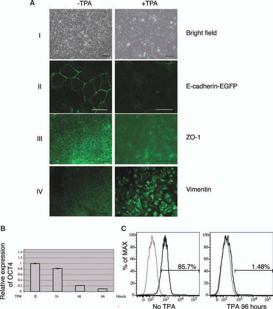
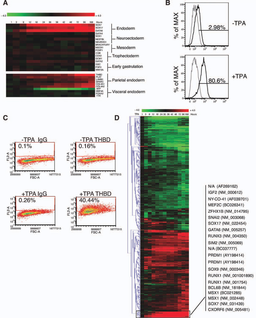
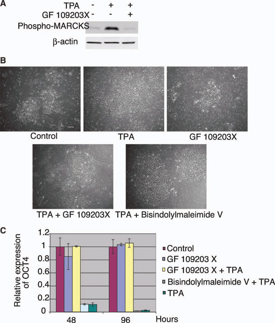
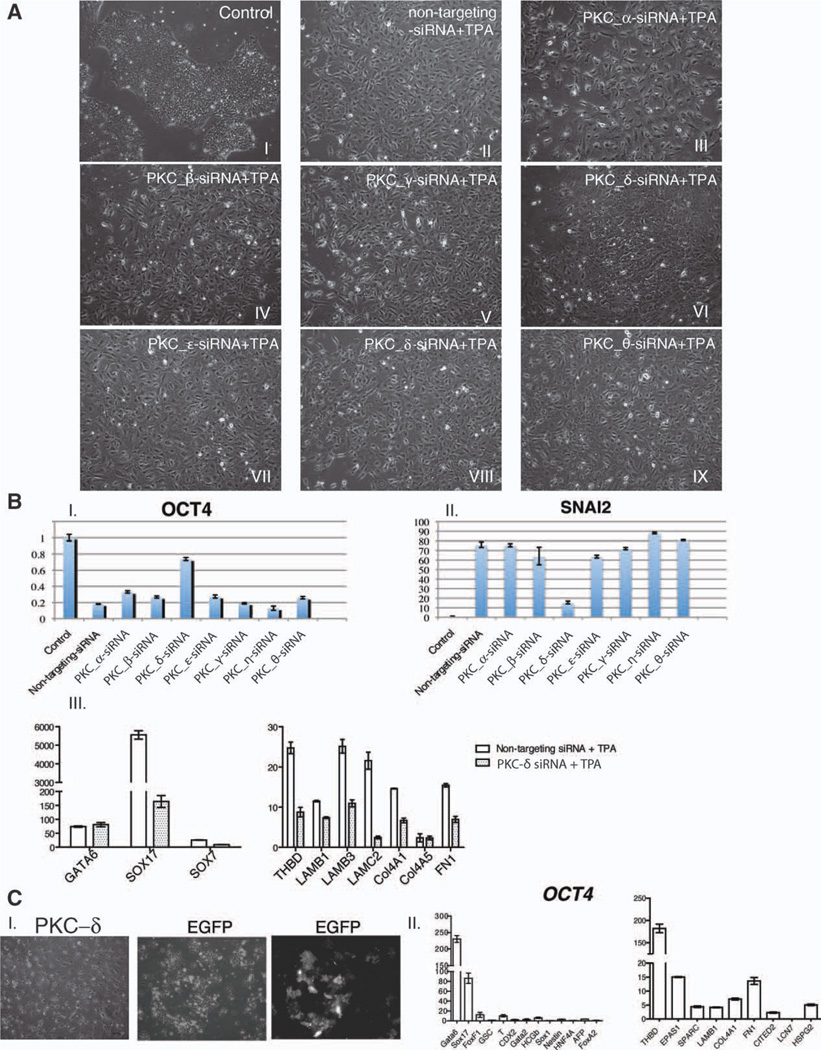
Unlike mouse embryonic stem cells (ESCs), which are closely related to the inner cell mass, human ESCs appear to be more closely related to the later primitive ectoderm. For example, human ESCs and primitive ectoderm share a common epithelial morphology, growth factor requirements, and the potential to differentiate to all three embryonic germ layers. However, it has previously been shown that human ESCs can also differentiate to cells expressing markers of trophoblast, an extraembryonic lineage formed before the formation of primitive ectoderm. Here, we show that phorbol ester 12-O-tetradecanoylphorbol 13-acetate causes human ESCs to undergo an epithelial mesenchymal transition and to differentiate into cells expressing markers of parietal endoderm, another extraembryonic lineage. We further confirmed that this differentiation is through the activation of protein kinase C (PKC) pathway and demonstrated that a particular PKC subtype, PKC-δ, is most responsible for this transition.
Copyright © 2011 AlphaMed Press.
Conflict of interest statement
The authors do not declare any conflicts of interest.
Figures
Similar articles
-
Induction and selection of Sox17-expressing endoderm cells generated from murine embryonic stem cells.Cells Tissues Organs. 2012;195(6):507-23. doi: 10.1159/000329864. Epub 2011 Nov 25. Cells Tissues Organs. 2012. PMID: 22123608
-
Stk40 links the pluripotency factor Oct4 to the Erk/MAPK pathway and controls extraembryonic endoderm differentiation.Proc Natl Acad Sci U S A. 2010 Jan 26;107(4):1402-7. doi: 10.1073/pnas.0905657107. Epub 2010 Jan 4. Proc Natl Acad Sci U S A. 2010. PMID: 20080709 Free PMC article.
-
Retinoic acid-induced transition from protein kinase C beta to protein kinase C alpha in differentiated F9 cells: correlation with altered regulation of proto-oncogene expression by phorbol esters.Cell Growth Differ. 1996 May;7(5):595-602. Cell Growth Differ. 1996. PMID: 8732669
-
Extraembryonic endoderm cells as a model of endoderm development.Dev Growth Differ. 2013 Apr;55(3):301-8. doi: 10.1111/dgd.12036. Epub 2013 Feb 18. Dev Growth Differ. 2013. PMID: 23414197 Review.
-
Differentiation of definitive endoderm from mouse embryonic stem cells.Results Probl Cell Differ. 2012;55:303-19. doi: 10.1007/978-3-642-30406-4_17. Results Probl Cell Differ. 2012. PMID: 22918814 Review.
Cited by
-
12-O-Tetradecanoylphorbol-13-acetate increases cardiomyogenesis through PKC/ERK signaling.Sci Rep. 2020 Sep 28;10(1):15922. doi: 10.1038/s41598-020-73074-4. Sci Rep. 2020. PMID: 32985604 Free PMC article.
-
Protein kinase C regulates human pluripotent stem cell self-renewal.PLoS One. 2013;8(1):e54122. doi: 10.1371/journal.pone.0054122. Epub 2013 Jan 21. PLoS One. 2013. PMID: 23349801 Free PMC article.
-
Protein kinases and associated pathways in pluripotent state and lineage differentiation.Curr Stem Cell Res Ther. 2014;9(5):366-87. doi: 10.2174/1574888x09666140616130217. Curr Stem Cell Res Ther. 2014. PMID: 24998240 Free PMC article. Review.
-
Signaling networks in human pluripotent stem cells.Curr Opin Cell Biol. 2013 Apr;25(2):241-6. doi: 10.1016/j.ceb.2012.09.005. Epub 2012 Oct 22. Curr Opin Cell Biol. 2013. PMID: 23092754 Free PMC article. Review.
-
Absence of catalytic domain in a putative protein kinase C (PkcA) suppresses tip dominance in Dictyostelium discoideum.Dev Biol. 2015 Sep 1;405(1):10-20. doi: 10.1016/j.ydbio.2015.05.021. Epub 2015 Jul 13. Dev Biol. 2015. PMID: 26183108 Free PMC article.
References
-
- Brons IG, Smithers LE, Trotter MW, et al. Derivation of pluripotent epiblast stem cells from mammalian embryos. Nature. 2007;448:191–195. - PubMed
-
- Tesar PJ, Chenoweth JG, Brook FA, et al. New cell lines from mouse epiblast share defining features with human embryonic stem cells. Nature. 2007;448:196–199. - PubMed
-
- Thomson JA, Itskovitz-Eldor J, Shapiro SS, et al. Embryonic stem cell lines derived from human blastocysts. Science. 1998;282:1145–1147. - PubMed
-
- Toyooka Y, Shimosato D, Murakami K, et al. Identification and characterization of subpopulations in undifferentiated ES cell culture. Development. 2008;135:909–918. - PubMed
-
- Nichols J, Chambers I, Taga T, et al. Physiological rationale for responsiveness of mouse embryonic stem cells to gp130 cytokines. Development. 2001;128:2333–2339. - PubMed
Publication types
MeSH terms
Substances
Grants and funding
LinkOut - more resources
Full Text Sources
Molecular Biology Databases
