. 2012 Jul;30(7):1095-102.
doi: 10.1002/jor.22038. Epub 2011 Dec 28.

Evaluation of BMP-2 gene-activated muscle grafts for cranial defect repair

Affiliations

Evaluation of BMP-2 gene-activated muscle grafts for cranial defect repair

Fangjun Liu et al. J Orthop Res. 2012 Jul.

Abstract

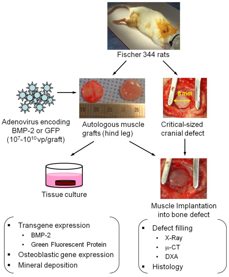
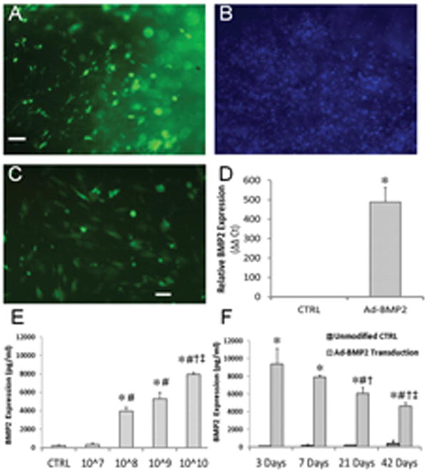
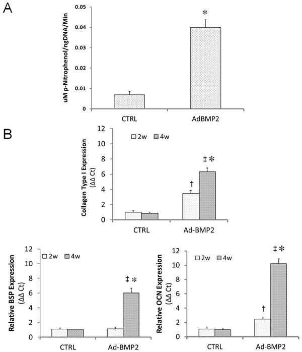
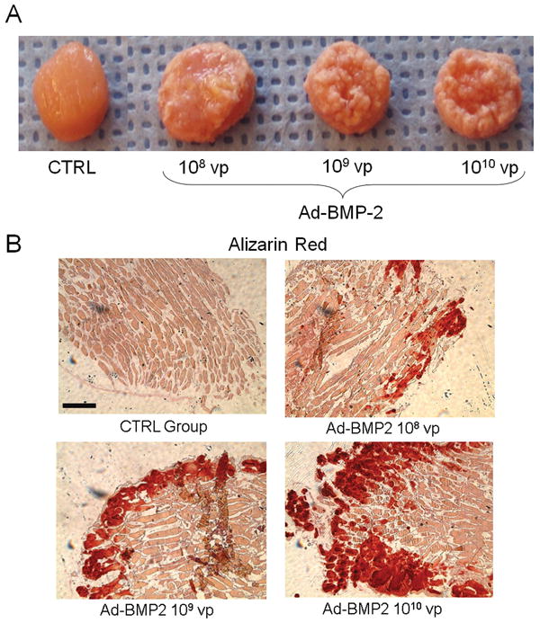
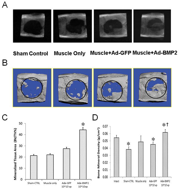
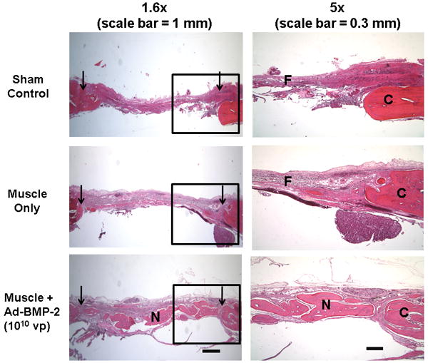
Large, osseous, segmental defects heal poorly. Muscle has a propensity to form bone when exposed to an osteogenic stimulus such as that provided by transfer and expression of cDNA encoding bone morphogenetic protein-2 (BMP-2). The present study evaluated the ability of genetically modified, autologous muscle to heal large cranial defects in rats. Autologous grafts (8 mm × 2 mm) were punched from the biceps femoris muscle and transduced intraoperatively with recombinant adenovirus vector containing human BMP-2 or green fluorescent protein cDNA. While the muscle biopsies were incubating with the vector, a central parietal 8 mm defect was surgically created in the calvarium of the same animal. The gene-activated muscle graft was then implanted into the cranial defect. After 8 weeks, crania were examined radiographically, histologically, and by micro-computed tomography and dual energy X-ray absorptiometry. Although none of the defects were completely healed in this time, muscle grafts expressing BMP-2 deposited more than twice as much new bone as controls. Histology confirmed the anatomical integrity of the newly formed bone, which was comparable in thickness and mineral density to the original cranial bone. This study confirms the in vivo osteogenic properties of genetically modified muscle and suggests novel strategies for healing bone.


Figures

Figure 1
Figure 1. Experimental design
Skeletal muscle was harvested from the hind limbs of Fischer F344 rats, and 8 mm discs were made using a biopsy punch. Discs were transduced with adenoviral vectors encoding human BMP-2 or GFP and cultured in vitro to evaluate transgene expression and consequent osteogenic response. To determine whether gene activated muscle could improve cranial bone healing, critical size defects were generated in rat skulls. Test groups received autologous muscle graft with or without intraoperative adenoviral modification. After 8 weeks, defect healing was evaluated qualitatively by X-Ray imaging and histology as well as quantitatively by μ-CT and DXA.
Figure 2
Figure 2. Transgene expression within genetically-modified muscle discs
(A-C) Fluorescence microscopic evaluation of muscle discs modified with Ad.GFP. The number of green fluorescent cells near the disc surface after 3 days (A) represents a considerable fraction of the total cell population, as indicated by DAPI staining (B). (C) Additional discs imaged 2 weeks post-modification demonstrate persistent GFP expression within the discs. Scale bars = 100 μm. (D) Discs modified with Ad.BMP-2 (1010 vp/disc) were cultured for 2 weeks, at which point BMP-2 message was detected by qRT-PCR. * indicates a significant increase (p<0.01) over unmodified muscle. (E) Conditioned media were collected from muscle discs transduced with 107 – 1010 vp Ad.BMP-2 after 1 week of culture, and BMP-2 protein levels were measured by ELISA. * and # indicate significant increases (p<0.01) over the control and 107 vp groups, respectively; † and ‡ represent significant increases (p<0.05) relative to the 108 and 109 vp groups, respectively. (F) BMP-2 levels within muscle-conditioned media at different time points following transduction with 1010 vp/disc Ad.BMP-2. * indicates a significant increase (p<0.01) over the control group; #, †, and ‡ represent significant decreases (p<0.05) relative to 3, 7, and 21 days, respectively.
Figure 3
Figure 3. Expression of osteoblastic markers by genetically-modified muscle discs
Discs were modified by 1010 vp Ad.BMP-2 and cultured for up to 4 weeks in basal osteogenic medium. (A) After 10 days in culture, alkaline phosphatase activity within muscle discs was measured and normalized by total DNA content. * indicates a significant increase (p<0.01) over the control group. (B) Additional discs were analyzed after 2 and 4 weeks of culture for expression of collagen type I, bone sialoprotein (BSP), and osteocalcin (OCN) by quantitative RT-PCR. † and ‡ denote significant increases (p<0.05) for the Ad.BMP-2 group relative to unmodified controls 2 and 4 weeks, respectively, while * indicates a significant difference (p<0.05) between the two time points for Ad.BMP-2-modified discs.
Figure 4
Figure 4. Mineral deposition within muscle grafts cultured in vitro
Muscle discs were modified with graded amounts of Ad.BMP-2 and cultured in basal osteogenic medium. (A) Gross appearance of muscle discs after 8 weeks. As the dose of Ad.BMP-2 and consequent BMP-2 secretion (see figure 2) increased, the discs became more extensively encased in a mineralized crust. (B) Alizarin red staining of disc cross sections revealed an increasingly thick layer of calcified mineral on the disc surface with increasing viral dose. Scale bar = 500 μm.
Figure 5
Figure 5. Cranial defect healing using gene-activated muscle grafts
Critical size defects were left empty (sham control), were filled with autologous, unmodified muscle (muscle control), or were filled with muscle modified intraoperatively with Ad.GFP or Ad.BMP-2 (1010 vp/disc). (A) Representative radiographs of skulls taken after 8 weeks. (B) Micro-computed tomography (μCT) scans reinforced observations from X-Ray imaging. (C) Ratios of bone volume to total volume were quantified from CT scans. * indicates a significant increase (p<0.05) for the Ad.BMP-2 group compared to Ad.GFP control. (D) Bone mineral density of the repair tissue was determined by DXA for the treatment groups as well as intact crania. * denotes a significant difference from the intact cranial bone (p<0.05); † indicates a significant difference compared to the Ad.BMP-2-muscle group.
Figure 6
Figure 6. Histological evaluation of filling within cranial defects
Transverse sections of decalcified rat crania were stained with hematoxylin and eosin (H&E). Left-hand column: images from representative sections of each experimental group are shown over the entire length of the defect. Arrowheads indicate the original defect boundaries. Scalebar = 1 mm. Right-hand column: higher-magnification regions (boxed areas from the left) are shown to better display one edge of the defect. “C” marks the original cranial bone, “N” denotes new bone formation within the defects, and “F” indicates fibrous repair tissue. Scale bar = 300 μm.

References

    1. Evans CH. Gene therapy for bone healing. Expert Rev Mol Med. 2010 Jun;12:e18. doi: 10.1017/S1462399410001493. - DOI - PMC - PubMed
    1. Evans CH, et al. Facilitated endogenous repair: making tissue engineering simple, practical, and economical. Tissue Eng. 2007;13(8):1987–93. - PubMed
    1. Shore EM, Kaplan FS. Insights from a rare genetic disorder of extra-skeletal bone formation, fibrodysplasia ossificans progressiva (FOP) Bone. 2008;43(3):427–33. - PMC - PubMed
    1. Stoltny T, et al. Heterotopic ossification in patients after total hip replacement. Ortop Traumatol Rehabil. 2007;9(3):264–72. - PubMed
    1. Covey DC. Combat orthopaedics: a view from the trenches. J Am Acad Orthop Surg. 2006;14(10 Spec No):S10–7. - PubMed

Publication types

Substances