Review
. 2012 May;113(5):1460-9.
doi: 10.1002/jcb.24046.

Medical therapies with adult stem/progenitor cells (MSCs): a backward journey from dramatic results in vivo to the cellular and molecular explanations

Affiliations
Review

Medical therapies with adult stem/progenitor cells (MSCs): a backward journey from dramatic results in vivo to the cellular and molecular explanations

Darwin J Prockop et al. J Cell Biochem. 2012 May.

Abstract

There is currently great interest in the use of mesenchymal stem/stromal cells (MSCs) for the therapy of many diseases of animals and humans. However, we are still left with the serious challenges in explaining the beneficial effects of the cells. Hence, it is essential to work backward from dramatic results obtained in vivo to the cellular and molecular explanations in order to discover the secrets of MSCs. This review will focus on recent data that have changed the paradigms for understanding the therapeutic potentials of MSCs.


Figures

Figure 1
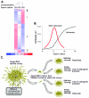
Figure 1. Summary of some of the unusual properties of MSCs in culture
(A) Heat map representation of Affymetrix microarray analysis of MSCs in culture at the log phase (at day 5, column D5), late log phase (at day 10, column D10), and stationary phase of growth (at day 15, column D15). Only the most differentially expressed genes between the conditions are represented, and they appear on the heat map as single colored segments arranged within each column. The color coding represents gene expression, where red is the highest amount of expression and blue is the lowest. Red segments represent gene expression that is 3 standard deviations over the mean of the three conditions (D5, D10, and D15), whereas blue segments represent gene expression that is 3 standard deviations under the mean of the three conditions. White segments indicate that the gene expression values are equal to the mean value of the three conditions, and lighter shades of red or blue represent less than 3 standard deviations above or below the mean, depending on the color intensity (see legend below map). Log-phase MSCs express the greatest number of cell cycle– and dedifferentiation-related genes (cluster labeled D), whereas more confluent, stationary-phase MSCs express genes related to conditioning the microenvironment, such as proteins of the extracellular matrix, cell surface adhesion molecules, and developmentally related genes (clusters labeled A to C). (B) The Wnt-inhibitor Dkk-1 is transiently expressed during the rapidly proliferating phase of MSC growth, probably to inhibit inappropriate differentiation. (C) A model to explain conditioning of MSCs by “microenvironmental niches” in vitro. MSCs are exposed to different cell densities within a single cell–derived colony (left). The heritable conditioning that occurs depends on the position of a given cell within the colony. When transferred to a new culture (center), the clonally derived MSCs behave differently in response to a given cocktail of soluble factors because of their microenvironmental preconditioning (right). The example here is a representation of a study assaying differentiation into osteoblasts and adipocytes, but the phenomenon is likely to apply to differentiation into other tissues as well. PPARγ, peroxide proliferator-activated receptorγ. Reprinted with permission from the American Association for the Advancement of Science (Gregory et al., 2005).
Figure 2
Figure 2. Anti-inflammatory effects of hMSCs activated to secrete TSG-6 in a mouse model of myocardial infarction
(A) Schematic diagram. ① Human MSCs (hMSCs) injected intravenously were trapped in the lungs and activated to secrete TSG-6 (TNF-α stimulated gene/protein 6). ② The TSG-6 decreased the normal but excessive inflammatory response that damages the heart. ③ The TSG-6 probably further decreased proteolytic damage to the heart by inhibiting matrix metalloproteinases (MMPs). (B) Selected sections through heart. Each heart was cut from apex to base into over 400 sequential 5 μm sections. Every twentieth section is shown. Either hMSCs or hMSCs transduced with the scrambled siRNA (scr siRNA) decreased the size of myocardial infarction examined 3 weeks later. However, hMSCs with a siRNA knockdown of the TSG-6 gene (TSG-6 siRNA) had no effect on infarct size. Intravenous infusion of 100 μg of recombinant human (rh) TSG-6 immediately following the surgery and at 24 hour also decreased infarct size. Panel (a) reproduced with modifications and with permission from Elsevier (Fang et al., 2007). Panel (b) reprinted with permission from Elsevier (Lee et al., 2009).
Figure 3
Figure 3. Dose dependent effects of TSG-6 in reducing corneal inflammation and opacity
Sterile inflammation was produced in corneas of Lewis rats by brief exposure to 100% ethanol followed by mechanical debridement of the cornea and limbal epithelium that removed the stem cells located in the limbus. (A) Representative corneal photographs on day 3 post-injury demonstrated that TSG-6 suppressed development of corneal opacity after chemical injury in a dose-dependent manner. (B) The anti-inflammatory effects of TSG-6 were dose-dependent as reflected in clinical grade of corneal opacity and myeloperoxidase (MPO) concentration as a semi-quantitative assay of neutrophil infiltration. Values are mean ± SD; n=3 for each group. (C) Gelatin zymography of corneas for pro-MMP-9 and active MMP-9. (D) Total and active MMP-9 concentration in the cornea as assayed by ELISA. Values are mean + SD; n=5 for each group. Significant improvements were observed with dose of 0.002 μg but maximal effects were obtained with 2 μg. Reprinted with permission from National Academy of Sciences, USA (Oh et al., 2010).
Figure 4
Figure 4. The anti-inflammatory effects of hMSCs and TSG-6 in a mouse model of zymosan-induced peritonitis
① Zymosan activated NF-κB signaling in resident macrophages via toll-like receptor 2 (TLR2). ② Activation of the NF-κB signaling pathway increased the production of pro-inflammatory cytokines to initiate the cascade of pro-inflammatory cytokines that was amplified by mesothelial cells and other cells of the peritoneum. ③ The pro-inflammatory cytokines also activated the hMSCs to secrete TSG-6. ④ TSG-6 decreased TLR2/NF-κB signaling in the resident macrophages through a direct interaction with CD44 or in a complex with hyaluronan. The amplification of the pro-inflammatory signals by mesothelial cells to recruit neutrophils was modulated by a negative feedback loop introduced by hMSCs and TSG-6.Reprinted with permission from the American Society of Hematology (Choi et al., 2011).
Figure 5
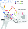
Figure 5. Schematic for the anti-inflammatory effects of MSCs based on observations in a mouse model for sepsis
Bacterial toxins such as LPS and circulating TNF-α acted on the TLR4 and TNF receptor-1 (TNFR-1) of MSCs to activate the NF-κB signaling. Activation of NF-κB signaling up-regulated expression of cyclooxygenase 2 (COX2) and the COX2 increased synthesis of prostaglandin E2 (PGE2). PGE2 was secreted and bound to EP2 and EP4 receptors on macrophages. The PGE2 thereby increased IL-10 secretion by macrophages to reduce the inflammatory response. Reprinted with permission from Macmillan Publishers Ltd (Németh et al., 2009).
Figure 6
Figure 6. Summary of some of the anti-inflammatory effects of MSCs
① Damage-associated molecular patterns (DAMPs) and IL-1α released by sterile injury or pathogen-associated molecular patterns (PAMPs) released by infectious injury to tissues activate resident macrophages through receptors involving pattern recognition receptors (PRRs). ② The activated macrophages produce pro-inflammatory cytokines such as IL-1α, IL-1β, or TNF-α to initiate the inflammatory cascade. ③ Simultaneously, the pro-inflammatory cytokines and probably other signals from injured cells activate MSCs to secrete anti-inflammatory factors that include TSG-6, PGE2, and IL-1ra that either modulate the activation of the resident macrophages or decrease the downstream effects of the pro-inflammatory cytokines. ④ The net effect is to decrease the amplification of the pro-inflammatory signals from resident macrophages by parenchymal cells through the secretion of IL-6, CXCL1, and related factors and to decrease the recruitment of neutrophils. Reprinted with permission from Macmillan Publishers Ltd (Prockop and Oh, 2011).
Figure 7
Figure 7
Schematic of how paradigms for therapies with MSCs have evolved.

References

    1. Ben-Ami E, Berrih-Aknin S, Miller A. Mesenchymal stem cells as an immunomodulatory therapeutic strategy for autoimmune diseases. Autoimmun Rev. 2011;10:410–415. - PubMed
    1. Bernardo ME, Locatelli F, Fibbe WE. Mesenchymal stromal cells. Ann N Y Acad Sci. 2009;1176:101–117. - PubMed
    1. Bianco P, Sacchetti B, Riminucci M. Osteoprogenitors and the hematopoietic microenvironment. Best Pract Res Clin Haematol. 2011;24:37–47. - PubMed
    1. Block GJ, Ohkouchi S, Fung F, Frenkel J, Gregory C, Pochampally R, DiMattia G, Sullivan DE, Prockop DJ. Multipotent stromal cells are activated to reduce apoptosis in part by upregulation and secretion of stanniocalcin-1. Stem Cells. 2009;27:670–681. - PMC - PubMed
    1. Caplan AI. Mesenchymal stem cells. J Orthop Res. 1991;9:641–650. - PubMed

Publication types

LinkOut - more resources