. 2012 Apr;81(4):343-50.
doi: 10.1002/cyto.a.22012. Epub 2011 Dec 29.

A novel Ly6C/Ly6G-based strategy to analyze the mouse splenic myeloid compartment

Affiliations

A novel Ly6C/Ly6G-based strategy to analyze the mouse splenic myeloid compartment

Shawn Rose et al. Cytometry A. 2012 Apr.

Abstract

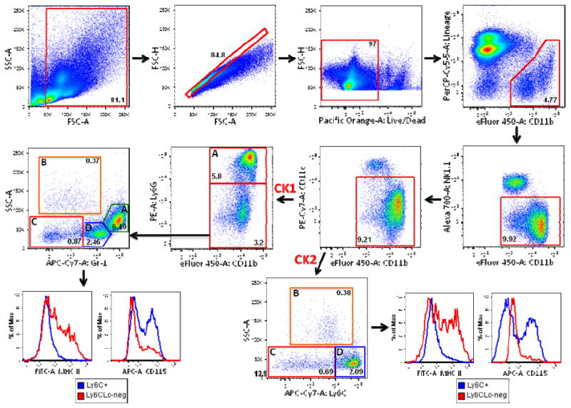
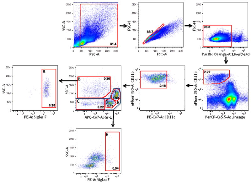
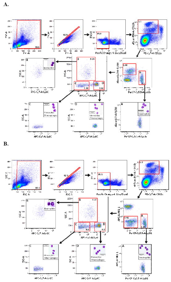
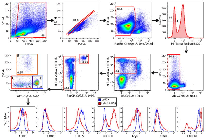
Currently, there is no standardized panel for immunophenotyping myeloid cells in mouse spleen using flow cytometry. Markers such as CD11b, CD11c, F4/80, Gr-1, Ly6C, and Ly6G have long been used to identify various splenic cell myeloid populations. Flow cytometry and fluorescence-activated cell sorting (FACS) analysis demonstrated that Ly6G/Ly6C markers are superior to Gr-1 for identifying splenic neutrophils, eosinophils, and subsets of monocytes/macrophages. Moreover, these experiments showed that F4/80 is not required for identifying these myeloid subsets and that many of the commercially available preparations of anti-F4/80 antibodies stain poorly for this antigen in spleen. Taken together, we have now developed an informative flow cytometry panel that can be combined with other cell markers to further delineate subpopulations of mouse splenic myeloid cells. This panel will be highly useful to investigators in the flow cytometry field, as there is a critical need to standardize the analysis of myeloid cell subsets.


Figures

Figure 1
Figure 1. Gr-1 and Ly6C/Ly6G markers identify similar populations of monocytes and macrophages in the mouse spleen
Total mouse splenocytes from C57BL/6 mice were prepared as described in Materials and Methods. Debris (SSC-A vs. FSC-A) and doublets (FSC-H vs. FSC-A) were excluded and live/dead discrimination was determined using the amine reactive dye Aqua (FSC-H vs. Pacific Orange-A Live/Dead). CD11b+Lineage- cells (gated out using a CD4, CD8, CD19 PerCP-Cy5.5 dump channel) were then sub-gated on CD11b+NK1.1- cells, followed by excluding CD11cHi cells. For the first cocktail (CK1), 4 subpopulations were identified: A) Gr-1HiSSCInt cells (neutrophils) and B) Gr-1Lo-negSSCHi cells (eosinophils) were negative for MHC Class II and CD115 staining (Supplemental Figure 5) C) Gr-1Lo-negSSCLo cells (monocytes/macrophages) were MHC Class II+/-CD115- (red histogram) D) Gr-1+SSCLo cells (monocytes/macrophages) were MHC Class II-CD115+/- (blue histogram). For the second cocktail (CK2), 4 subpopulations were identified: A) Ly6GHiCD11b+ cells (neutrophils) and B) Ly6CLo-negLy6G-SSCHi cells (eosinophils) were negative for MHC Class II and CD115 (Supplemental Figure 5) C) Ly6CLo-negLy6G-SSCLo cells (monocytes/macrophages) were MHCII+/-CD115- (red histogram) D) Ly6C+Ly6G-SSCLo cells (monocytes/macrophages) were MHC Class II-CD115+/- (blue histogram). Frequencies of cells in each sub-gate (after debris and doublet exclusion) are expressed as a percentage of live cells.
Figure 2
Figure 2. Gr-1Lo-negSSCHi mouse splenocytes are eosinophils
As in Figure 1, debris, doublets, and nonviable cells were excluded from total C57BL/6 mouse splenocytes. Live cells were sub-gated on CD11b+Lineage- cells (gated out using a CD4, CD8, CD19, NK1.1 PerCP-Cy5.5 dump channel). After gating out CD11cHi cells, 4 subpopulations of cells were identified: A) Gr-1HiSSCInt cells (neutrophils) B) Gr-1Lo-negSSCHi cells (eosinophils) were positive for Siglec F staining on sub-gate analysis C) Gr-1Lo-negSSCLo and D) Gr-1+SSCLo monocytes/macrophages were also identified. Gate E, which encompasses gates A, C, and D, was negative for Siglec F staining. Frequencies of cells in each sub-gate (after debris and doublet exclusion) are expressed as a percentage of live cells.
Figure 3
Figure 3. Monocytes/macrophages in the mouse spleen are readily identified without using the F4/80 antigen
A) Debris, doublets, and dead cells were excluded from total C57BL/6 mouse splenocytes as in Figures 1-2. CD11cHi cells were excluded from the CD11b+ population, followed by gating into F4/80+Ly6G- and F4/80-Ly6GHi (A) subpopulations. F4/80+Ly6G- cells were divided further into subpopulations B, C, and D based on Ly6C versus SSC. Populations A-D were then isolated by FACS. Cytospin preparations from the sorted cells were stained with Diff-Quick and Giemsa. Subpopulation A (F4/80-Ly6GHi) contained solely neutrophils. Subpopulation B (Ly6CLo-negSSCHi) contained eosinophils while subpopulations C (Ly6CLo-negSSCLo) and D (Ly6C+SSCLo) contained monocytes/macrophages. B) Cells were prepared and gated as in Figure 3A except that NK1.1 was substituted for F4/80 staining to distinguish NK1.1-Ly6G- and NK1.1-Ly6GHi (A) subpopulations. NK1.1-Ly6G- cells were divided into subpopulations B-D based on Ly6C versus SSC. Subpopulations A-D were then sorted and cytospin preparations were prepared as above. Subpopulation A (Ly6GHiNK1.1-) contained exclusively neutrophils. Subpopulation B (Ly6CLo-negSSCHi) contained eosinophils while subpopulations C (Ly6CLo-negSSCLo) and D (Ly6C+SSCLo) contained monocytes/macrophages. For all analyses, frequencies of cells in each sub-gate (after debris and doublet exclusion) are expressed as a percentage of live cells.
Figure 4
Figure 4. Immunophenotyping monocyte/macrophage subsets in mouse spleen
Debris, doublets, and dead cells were excluded from mouse splenocytes of CX3CR1-GFP/+ mice as described in Figure 1. B220+ cells were gated out followed by exclusion of NK1.1+ cells. CD11cHi cells were then gated out from the CD11b+ population and the remaining CD11b+ cells were separated into Ly6G- and (A) Ly6GHi (neutrophils) subpopulations. Ly6G- cells were divided further into subpopulations B (eosinophils), C, and D based on their Ly6C versus SSC properties. Monocyte/macrophage subpopulations C (red histograms) and D (blue histograms) were then further characterized for expression of various cell surface markers including CD80, CD86, CD115, MHC Class II (MHC II), FcγR, CD40, and CX3CR1 as indicated.

References

    1. Ziegler-Heitbrock L, Ancuta P, Crowe S, Dalod M, Grau V, Hart DN, Leenen PJ, Liu YJ, MacPherson G, Randolph GJ, et al. Nomenclature of monocytes and dendritic cells in blood. Blood. 2010;116:e74–80. - PubMed
    1. Yona S, Jung S. Monocytes: subsets, origins, fates and functions. Current Opinion in Hematology. 2010;17:53–9. - PubMed
    1. Mosser DM, Edwards JP. Exploring the full spectrum of macrophage activation. Nat Rev Immunol. 2008;8:958–69. - PMC - PubMed
    1. Austyn JM, Gordon S. F4/80, a monoclonal antibody directed specifically against the mouse macrophage. European Journal of Immunology. 1981;11:805–15. - PubMed
    1. Schaller E, Macfarlane AJ, Rupec RA, Gordon S, McKnight AJ, Pfeffer K. Inactivation of the F4/80 glycoprotein in the mouse germ line. Molecular and Cellular Biology. 2002;22:8035–43. - PMC - PubMed

Publication types