. 2012 Jan 9;196(1):65-83.
doi: 10.1083/jcb.201106088. Epub 2012 Jan 2.

dEHBP1 controls exocytosis and recycling of Delta during asymmetric divisions

Affiliations

dEHBP1 controls exocytosis and recycling of Delta during asymmetric divisions

Nikolaos Giagtzoglou et al. J Cell Biol. .

Abstract

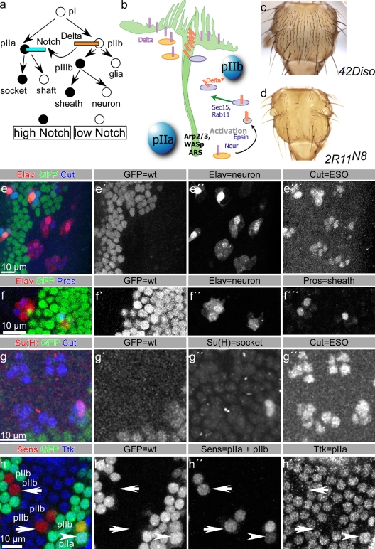
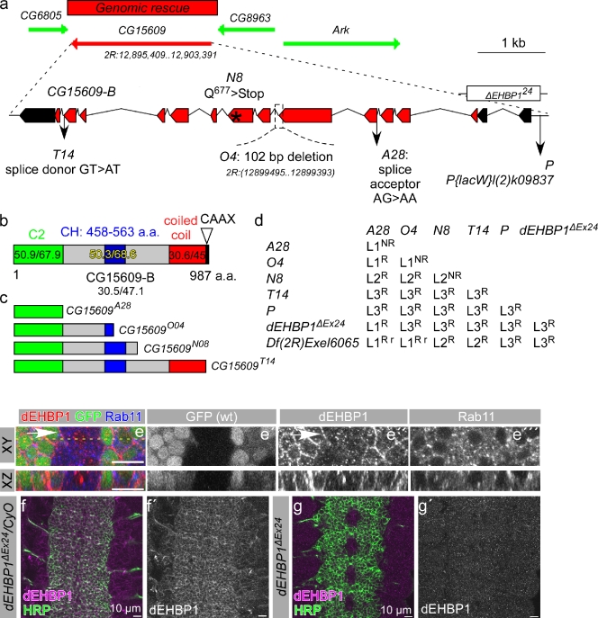
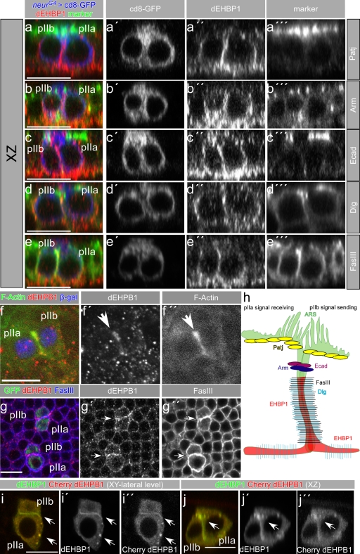
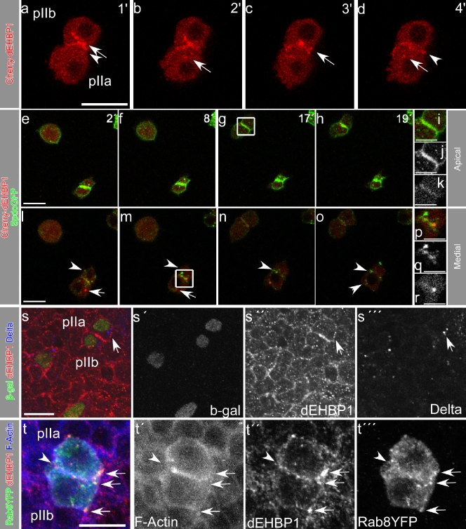
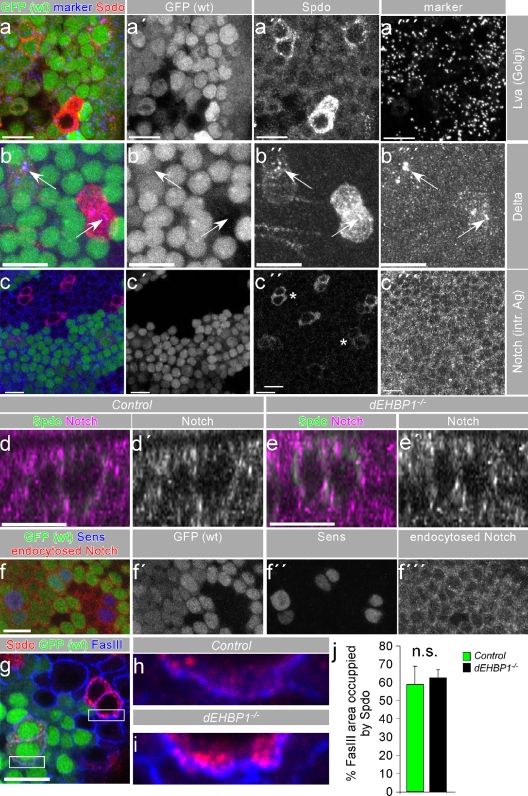
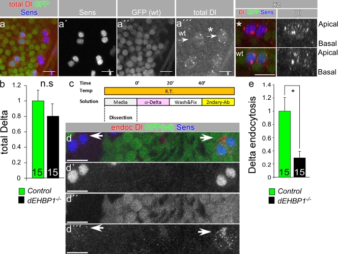
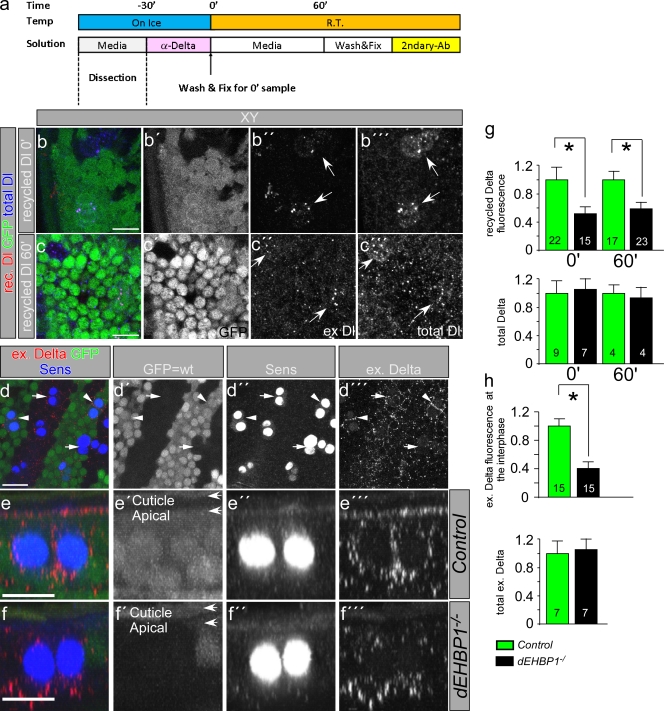
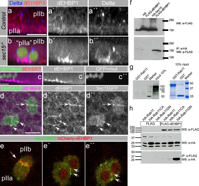
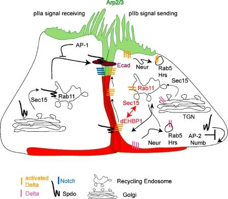
Notch signaling governs binary cell fate determination in asymmetrically dividing cells. Through a forward genetic screen we identified the fly homologue of Eps15 homology domain containing protein-binding protein 1 (dEHBP1) as a novel regulator of Notch signaling in asymmetrically dividing cells. dEHBP1 is enriched basally and at the actin-rich interface of pII cells of the external mechanosensory organs, where Notch signaling occurs. Loss of function of dEHBP1 leads to up-regulation of Sanpodo, a regulator of Notch signaling, and aberrant trafficking of the Notch ligand, Delta. Furthermore, Sec15 and Rab11, which have been previously shown to regulate the localization of Delta, physically interact with dEHBP1. We propose that dEHBP1 functions as an adaptor molecule for the exocytosis and recycling of Delta, thereby affecting cell fate decisions in asymmetrically dividing cells.


Figures

Figure 1.
Figure 1.
2R11 alleles disrupt Notch signaling in the asymmetrically dividing thoracic ESO lineages. (a) Diagram of the asymmetric divisions during development of the ESO lineage; black circles represent Notch signal–receiving cells, white circles represent Notch signal–sending cells. (b) A possible model for Notch signaling in asymmetrically dividing ESO lineages (adopted by Rajan et al., 2009). (c and d) Thoracic y−/− clones of the parental 42Diso chromosome (c) or of the 2R11N8 allele (generated in a Minute background) (d). (e–g′′′) Analysis of different cell type markers of the ESO lineage at 24 h APF; pupal thoraces reveal that mutant ESO cells acquire erroneous cell fates. (e–e′′′) Supernumerary, elav-positive neurons arise in 2R11 negatively marked clones. (f–f′′′) Extra prospero–positive sheath and elav-positive neuron cells develop in 2R11 thoracic clones. (g–g′′′) Su(H)-positive socket cells are absent from 2R11 clones. In e-g′′′, cells of the ESO lineages are marked by Cut. (h–h′′′) Tramtrack-positive pIIa cells are absent from dEHBP1A28 clones within pupal nota at 17 h APF, revealing that Notch signaling is affected within the mutant regions. pIIa and pIIb cells are stained for Sens. Arrows indicate mutant pIIa cells, and the arrowhead points to a wild-type pIIa cell. The alleles used in e–e′′′ and g–g′′′ are dEHBP1O4. The alleles used in f–f′′′ and h–h′′′ are dEHBP1A28. Bars, 10 µm.
Figure 2.
Figure 2.
2R11 alleles map to CG15609, the fly homologue of Eps15 homology domain containing protein-binding protein 1. (a) Diagram of the genomic locus of CG15609 (red arrow), the genomic rescue construct (red box), and the exon–intron structure of CG15609 isoform B (red bars signify coding exons, black bars signify non coding exons), where the molecular lesions of 2R11 alleles are shown. ΔEHBP1ex24 is a deletion caused by imprecise excision of P{lacW}l(2)k09837 (indicated as P allele). (b) Schematic representation of dEHBP1 protein structure. Percentages indicate identity/similarity between the fly and mouse homologues. C2 represents the lipid-binding domain and is colored in green, CH stands for calponin homology actin-binding domain and is colored in blue, coiled coil protein interaction domain is colored in red, and CAAX (C, cysteine; A, aliphatic; X, any amino acid) motif is shown as a triangle at the end of the protein sequence. a.a., amino acid. (c) The predicted structure of the dEHBP1 protein, in different mutant 2R11 alleles. (d) Lethal phase analysis of different mutations. NR, not rescued lethality; R, lethality rescued by genomic rescue; r, lethality rescued by cDNA, expressed ubiquitously by tubGal4 driver. (e–e′′′) Anti-dEHBP1 fails to recognize the majority of the protein in pupal thoracic homozygous clones of CG15609O4. Sections at both XY and XZ levels (indicated by yellow dashed line in e) are shown. Single-channel representations are shown in (e′) for GFP (wild-type region), (e′′) for dEHBP1, and (e′′′) for Rab11, which marks the recycling endosome. (f–g′) Anti-dEHBP1specifically recognizes dEHBP1 in embryonic CNS of control, balanced embryos (f–f′′) in comparison to CNS from homozygous mutant siblings (g–g′′). Bars, 10 µm.
Figure 3.
Figure 3.
dEHBP1 is enriched at the basolateral side of the plasma membrane and colocalizes with F-actin at the interface of pIIa and pIIb cells. (a–e′′′) Analysis of the subcellular localization of dEHBP1 along the z axis with respect to various markers of apico-basal polarity in ESO cells (marked by neurGal>UAS-cd8-GFP), such as Patj (a–a′′′), Arm (b–b′′′), E-cad (c–c′′′), Dlg (d–d′′′), and FasIII (e–e′′′). (f–f′′) dEHBP1 colocalizes with F-actin at the interface of pIIa and pIIb cells, marked by the nuclear β-galactosidase (β-gal) in a neurA101 enhancer trap fly strain. (g–g′′) dEHBP1 exhibits a punctate pattern in thoracic epithelia. Arrows point to the enrichment of dEHBP1 at the interface of ESO cells (marked by neurGal>UAS-cd8-GFP). Bars, 10 µm. (h) Diagram depicting the relative localization of dEHBP1 with respect to markers of apico-basal polarity in ESO cells. Only the most prominent, basolateral expression of dEHBP1 with emphasis to the interface of ESO cells is shown, for the sake of clarity. (i–j′′) mCherry-dEHBP1, expressed in ESO lineages by neurGal4, localizes at the interphase of pII cells and within intracellular punctae, also recognized by the anti-dEHBP1 antibody. Sections are shown at the xy (i–i′′) as well as at the xz level (j–j′′).
Figure 4.
Figure 4.
dEHBP1 is transiently enriched at the interface of the pIIa/pIIb cells, where it partially colocalizes with Spdo. (a–d) Still frames from Video 1 showing medial sections of pIIa/pIIb cells that contain mCherry-dEHBP1 in intracellular punctae, pointed by arrows, as well as the interface of the cells, indicated by arrowheads. Numbers at the top right corner indicate the minutes of live imaging. (e–k) Still frames from Videos 2 and 3 showing apical and medial sections of pIIa/pIIb cells, respectively, that express mCherry-dEHBP1 and Spdo-GFP, which colocalize along the interface of dividing pII cells toward the subapical regions (i–k). Numbers at the top right corner indicate the minutes of live imaging. (i–k) Magnification of the pIIa/pIIb cells included in the box in g. (l–r) mCherry-dEHBP1 (arrow) and Spdo GFP punctae (arrowheads) do not colocalize, but only at the interface of dividing pII cells (p–r). (p–r) Magnification of the pIIa/pIIb interface included in the box in m. Single-channel representations are shown in black and white for Spdo-GFP (q) and for mCherry-dEHBP1 (r). (s-s′′′) Single confocal sections of pIIa/pIIb cells of neurA101 strain, marked by nuclear β-gal, indicate that dEHBP1 does not colocalize with Delta punctae (arrow). (t–t′′′) Rab8YFP, expressed in pII cells by neurGal4, colocalizes with dEHBP1 in vesicular structures as well as at the actin-rich interphase of pII cells, as indicated by the arrows and arrowheads, respectively. Bars: (p–r) 5 µm, (all others) 10 µm.
Figure 5.
Figure 5.
Spdo is up-regulated in the absence of dEHBP1. (a–b′′′) Spdo is up-regulated in dEHBP1−/− ESO cells but not restricted in a Lva-positive Golgi compartment (a–a′′′) and not accompanied by a similar accumulation of Delta (b–b′′′). Arrows indicate to the colocalization of intense Spdo punctae with Delta in both wild-type and mutant cells. (c–c′′′) Spdo is up-regulated in dEHBP1−/− ESO cells, but not accompanied by accumulation of Notch (d–e′) Notch does not display aberrant localization along the xz axis in ESO clusters, indicated by the asterisks in c′′. (f–f′′′) Endocytosis of Notch is not altered in the absence of dEHBP1. (g–i) Spdo is up-regulated in dEHBP1−/− ESO cells, but not excluded from the FasIII-positive part of the plasma membrane, as shown in single confocal sections. Magnification of parts of the plasma membrane, included in the boxed regions in g, are shown for a wild-type, GFP-positive ESO cluster (h) and for mutant, GFP-negative, ESO clusters (i). (j) Quantification of the FasII-positive area that is occupied by Spdo in control and dEHBP1−/− ESO clusters indicates that Spdo is able to reach the plasma membrane in the absence of dEHBP1. Bars, 10 µm.
Figure 6.
Figure 6.
Delta endocytosis is impaired in the absence of dEHBP1. (a–a′′′) The total amount of Delta is not affected upon loss of dEHBP1. The asterisk marks the dEHBP1−/− cell cluster, and wt stands for the wild-type control cluster. (b) Normalized quantification of levels of total Delta in wild-type (green bars) and dEHPB1−/− (black bars) ESO clusters. Numbers at the base of the bars represent the number of ESO clusters used for quantification; n.s., not significant. (c) Schematic representation of the Delta endocytosis assays; R.T., room temperature. (d–d′′′) Delta endocytosis is impaired under conditions of loss of function of dEHBP1. Arrows point to the Delta punctae detected in the endocytosis assay. Few, small punctae are still seen in the mutant ESOs, suggesting that endocytosis is not completely abolished. (e) Normalized quantification of endocytosed Delta (in d–d′′′) in wild-type (green bars) and mutant (black bars) ESO two-cell clusters. Numbers at the base of the bars represent the number of ESOs of two-cell clusters used for quantification. *, P < 0.05.
Figure 7.
Figure 7.
Delta localization at the signaling interface of the pIIa/pIIb cells is impaired in the absence of dEHBP1. (a) Schematic representation of the Delta pulse-chase assays. R.T., room temperature. (b–c′′′) Delta is reduced in dEHBP1−/− pIIa and pIIb cells at 0-min time points (b–b′′′) and at the 60-min time point (c–c′′′) in pulse-chase assays, as shown in xy projections. In b′′, b′′′, c′′, and c′′′, arrows point to the interface of mutant and wild-type two-cell ESO clusters, respectively. (d–f′′′) Extracellular Delta is reduced in dEHBP1−/− pIIa and pIIb cells, as shown in xy projections in d–d′′′, where arrows and arrowheads point to the interface of mutant and wild-type two-cell ESO clusters, respectively. Analysis of extracellular Delta in projections along the z axis of wild-type thoracic (e–e′′′′) and mutant (f–f′′′) thoracic clusters, respectively, reveal that Delta is severely reduced at the interface of the pIIa and pIIb cells and that it is mainly localized basally. (g) Normalized quantification of pulse-chased and total Delta at 0- and 60-min time points in wild-type (green bars) and mutant (black bars) ESO two-cell clusters. (h) Normalized quantifications of extracellular Delta at the interphase of wild-type (green bars) and mutant (black bars) pII cells, as well as total extracellular Delta throughout wild-type (green bars) and mutant (black bars) thoracic epithelia. The numbers at base of the bars in g and h represent the total number of clusters used for quantification of Delta. n.s., not significant; *, P < 0.05. Bars, 10 µm.
Figure 8.
Figure 8.
dEHBP1 accumulates with Delta in the absence of Sec15 and it physically interacts with Sec15 and Rab11. (a–b′′′) xz projection of single ESO clusters within thoracic epithelia, which are wild-type (a–a′′) or homozygous mutant for sec15 (b–b′′, positively marked by the expression of GFP), reveals that dEHBP1 accumulates basally along with Delta in the absence of sec15. (c–c′′) XZ projection of thoracic epithelia, shown in a–b, which contain homozygous mutant cells for sec15 (c′′, positively marked by the expression of GFP), reveals that dEHBP1 accumulates basally in all epithelial cells in pupal thoraces in the absence of sec15. (d–d′′) Sec15-GFP, overexpressed by neurGal4, and dEHBP1 colocalize in ESO clusters at the interface of the progeny, as indicated by the arrows. Single-channel representations for dEHBP1 (d′) and Sec15-GFP (d′′) are shown in black and white. (e–e′′) Sec15-GFP and mCherry-dEHBP1 overexpressed by neurGal4 colocalize in ESO clusters. (f) HA-Sec15 and FLAG-dEHBP1 coimmunoprecipitate from whole-cell lysates of transiently transfected S2 cells. (g) GST-Rab11 does not interact with in vitro–translated dEHBP1. 10% input of the proteins used for the GST pull-down were analyzed by Coomassie stain to ensure the integrity and equal amounts of GST proteins. (h) dEHBP1 interacts weakly with wild-type and constitutively active Rab11 variants, but strongly with dominant-negative Rab11 in coIP experiments from S2 cells. Bars, 10 µm.
Figure 9.
Figure 9.
Model of dEHBP1 function. Diagram adopted from Rajan et al. (2009), depicting a revised version of Delta recycling pathway. Neur-mediated endocytosis of Delta occurs at both the basal and the apical sides of the pIIb cell and results in the delivery of Delta to Rab5 early endosomes (Benhra et al., 2011). A fraction of Delta then follows a Rab11–Sec15-dependent route (Emery et al., 2005; Jafar-Nejad et al., 2005; Benhra et al., 2011) toward the ARS (Rajan et al., 2009). dEHBP1 (represented by red region overlapping the green region marking the ARS) is enriched at the actin-rich interface of the asymmetrically dividing pII cells where it may facilitate the localization of Delta via its interaction with Sec15 and Rab11. AP-2 and Numb inhibit the localization of Spdo at the plasma membrane of the pIIb cell (O’Connor-Giles and Skeath, 2003; Hutterer and Knoblich, 2005), while AP-1 inhibits the recycling of Spdo toward the apical portion of the pIIa cell (Benhra et al., 2010). Spdo depends on Sec15 activity to reach the plasma membrane (Tong et al., 2010).

Similar articles

Cited by

References

    1. Acar M., Jafar-Nejad H., Takeuchi H., Rajan A., Ibrani D., Rana N.A., Pan H., Haltiwanger R.S., Bellen H.J. 2008. Rumi is a CAP10 domain glycosyltransferase that modifies Notch and is required for Notch signaling. Cell. 132:247–258 10.1016/j.cell.2007.12.016 - DOI - PMC - PubMed
    1. Andrews H.K., Giagtzoglou N., Yamamoto S., Schulze K.L., Bellen H.J. 2009. Sequoia regulates cell fate decisions in the external sensory organs of adult Drosophila. EMBO Rep. 10:636–641 10.1038/embor.2009.66 - DOI - PMC - PubMed
    1. Ang A.L., Fölsch H., Koivisto U.M., Pypaert M., Mellman I. 2003. The Rab8 GTPase selectively regulates AP-1B-dependent basolateral transport in polarized Madin-Darby canine kidney cells. J. Cell Biol. 163:339–350 10.1083/jcb.200307046 - DOI - PMC - PubMed
    1. Artavanis-Tsakonas S., Rand M.D., Lake R.J. 1999. Notch signaling: cell fate control and signal integration in development. Science. 284:770–776 10.1126/science.284.5415.770 - DOI - PubMed
    1. Babaoglan A.B., O’Connor-Giles K.M., Mistry H., Schickedanz A., Wilson B.A., Skeath J.B. 2009. Sanpodo: a context-dependent activator and inhibitor of Notch signaling during asymmetric divisions. Development. 136:4089–4098 10.1242/dev.040386 - DOI - PMC - PubMed

Publication types

MeSH terms

Substances