. 2012 Mar;63(5):2089-103.
doi: 10.1093/jxb/err414. Epub 2012 Jan 2.

S-Nitrosylated proteins in pea (Pisum sativum L.) leaf peroxisomes: changes under abiotic stress

Affiliations

S-Nitrosylated proteins in pea (Pisum sativum L.) leaf peroxisomes: changes under abiotic stress

Ana P Ortega-Galisteo et al. J Exp Bot. 2012 Mar.

Abstract

Peroxisomes, single-membrane-bounded organelles with essentially oxidative metabolism, are key in plant responses to abiotic and biotic stresses. Recently, the presence of nitric oxide (NO) described in peroxisomes opened the possibility of new cellular functions, as NO regulates diverse biological processes by directly modifying proteins. However, this mechanism has not yet been analysed in peroxisomes. This study assessed the presence of S-nitrosylation in pea-leaf peroxisomes, purified S-nitrosylated peroxisome proteins by immunoprecipitation, and identified the purified proteins by two different mass-spectrometry techniques (matrix-assisted laser desorption/ionization tandem time-of-flight and two-dimensional nano-liquid chromatography coupled to ion-trap tandem mass spectrometry). Six peroxisomal proteins were identified as putative targets of S-nitrosylation involved in photorespiration, β-oxidation, and reactive oxygen species detoxification. The activity of three of these proteins (catalase, glycolate oxidase, and malate dehydrogenase) is inhibited by NO donors. NO metabolism/S-nitrosylation and peroxisomes were analysed under two different types of abiotic stress, i.e. cadmium and 2,4-dichlorophenoxy acetic acid (2,4-D). Both types of stress reduced NO production in pea plants, and an increase in S-nitrosylation was observed in pea extracts under 2,4-D treatment while no total changes were observed in peroxisomes. However, the S-nitrosylation levels of catalase and glycolate oxidase changed under cadmium and 2,4-D treatments, suggesting that this post-translational modification could be involved in the regulation of H(2)O(2) level under abiotic stress.


Figures
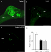
Fig. 1.
Fig. 1.
Nitric oxide (NO) production in pea leaves by confocal laser scanning fluorescence microscopy, after treatment with 22.6 mM 2,4-dichlorophenoxy acetic acid (2,4-D). (A–C) Maximum projections of several optical sections collected by confocal microscopy showing fluorescence due to 4,5-diaminoflorescein diacetate (excitation at 495 nm, emission at 520 nm) in leaf cross-sections of (A) control and (B) 2,4-D-treated plants; (C) negative control, leaves from control plants incubated with 300 μM carboxy 2-phenyl-4,4,5,5-tetramethylimidazoline-1-oxyl 3-oxide (cPTIO), which acts as NO scavenger. (D) Histogram showing relative fluorescence intensities corresponding to NO quantified in arbitrary units using LCS Lite software from Leica. Different letters indicate significant difference (P < 0.01) according to Student’s t-test. These results are representative of at least three independent experiments. c, collenchyma; mc, mesophyll cells; scl, sclerenchyma; x, xylem vessels. Bars, 250 μm.
Fig. 2.
Fig. 2.
Effect of 2,4-dichlorophenoxy acetic acid (2,4-D) on the activity of S-nitrosoglutathione reductase (GSNOR) in pea leaves. Each column represents the mean ± SE of three independent experiments. *, Differences were significant at P < 0.01 according to Student’s t-test.
Fig. 3.
Fig. 3.
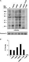
Detection of S-nitrosylated proteins in pea-leaf (Pisum sativum L.) extracts. (A) Protein extracts (150 μg) from pea leaves were not treated (control, C) or treated with 1 mM glutathione disulphide (GSSG), 1 mM S-nitroso-N-acetylpenicillamine (SNAP), 1 mM S-nitrosoglutathione (GSNO), or 10 mM tris(2-carboxyethyl)phosphine (TCEP) previously treated or not with GSNO and were subjected to the biotin-switch assay, separated by SDS-PAGE, and immunoblotted with an anti-biotin antibody. Protein loading was verified by Ponceau staining. (B) Histogram showing relative quantification of the Western blot showed in (A). The intensity of bands was quantified using Quantity One Software (version 4.6.2). Band intensity was expressed as fold-change (FC) density. Each band density was first normalized by dividing it by the density of the Ponceau band in the same lane. Then, the relative increase or decrease in density was calculated by dividing the normalized band density of the problem sample by the normalized band density of the control sample. The results are representative of three different Western blots assayed.
Fig. 4.
Fig. 4.
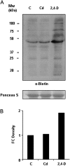
Detection of S-nitrosylated proteins in pea-leaf (Pisum sativum L.) extracts under abiotic stress. Protein extracts (150 μg) from pea leaves were not treated (control, C) or treated with 50 μM cadmium or 22.6 mM 2,4-dichlorophenoxy acetic acid (2,4-D) and were subjected to the biotin-switch assay, separated by SDS-PAGE, and immunoblotted with an anti-biotin antibody. Protein loading was verified by Ponceau staining. B, Histogram showing relative quantification of Western blot showed in (A). The intensity of bands was quantified as described in the legend for Fig. 3. The results are representative of four different Western blots assayed.
Fig. 5.
Fig. 5.
Subcellular localization of S-nitrosoglutathione (GSNO) and glutathione (GSH) in pea leaves. (A) Pre-immune serum control. (B) Pea-leaf cells with anti-GSNO 1:250. (C) Pea-leaf cells with anti-GSH 1:250. Immunogold labelling of GSH and GSNO are indicated by arrowheads. CL, chloroplast; M, mitochondrion; P, peroxisome; PC, cell wall. Bars, 1 μm.
Fig. 6.
Fig. 6.
Detection of S-nitrosylated proteins in pea-leaf (Pisum sativum L.) peroxisomes. Peroxisomal proteins (250 μg) from pea plants were not treated (control, C), treated with 50 μM cadmium or 22.6 mM 2,4-dichlorophenoxy acetic acid (2,4-D), or incubated with 1 mM S-nitrosoglutathione (C+GSNO) or with 20 mM dithiothreitol previously incubated with GSNO (C+GSNO+DTT) and were subjected to the biotin-switch assay, separated by SDS-PAGE, and immunoblotted with an anti-biotin antibody. Protein loading was verified by Ponceau staining of the membrane. The experiment was repeated three times with similar results.
Fig. 7.
Fig. 7.
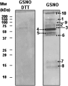
S-Nitrosylated proteins of peroxisomes from pea leaves (Pisum sativum L.). Peroxisomal proteins (5 mg) were treated with 1 mM S-nitrosoglutathione (GSNO) and then with 20 mM dithiothreitol (DTT) or not and were subjected to the biotin-switch assay. Biotinylated proteins were purified by immunoprecipitation with an anti-biotin antibody. Eluates were separated by SDS-PAGE and stained by Coomassie blue. The protein bands were identified by matrix-assisted laser desorption/ionization tandem time-of-flight (Table 1).
Fig. 8.
Fig. 8.
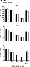
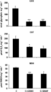
Effect of NO on glycolate oxidase (GOX), catalase (CAT), and malate dehydrogenase (MDH) activities. Commercial proteins were preincubated with different concentrations (0–1000 μM) of GSNO (black bars) or diethylenetriamine (DETA) NONOate (white bars) for 30 min at room temperature and then enzyme activities were determined (Materials and methods). Incubation with 10 mM dithiothreitol (DTT) after 1000 μM GSNO or DETA NONOate restored enzyme activities. For each concentration, measurements were made in triplicate. Different letters indicate significant differences (P < 0.001 for GOX and MDH and P < 0.01 for CAT), as determined by Student’s t-test.
Fig. 9.
Fig. 9.
Effect of NO on glycolate oxidase (GOX), catalase (CAT), and malate dehydrogenase (MDH) activities in pea leaves (Pisum sativum L.). Pea extracts for measuring CAT and GOX activity and pea peroxisomes for MDH activity were preincubated with 1 mM S-nitrosoglutathione (GSNO) and 1 mM S-nitroso-N-acetylpenicillamine (SNAP) for 45 min at room temperature and then enzyme activities were determined (Materials and methods). Three different extracts were used for each measurement. Asterisk indicates significant differences (*, P < 0.05; ***, P < 0.001) according to Student’s t-test.
Fig. 10.
Fig. 10.
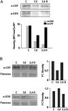
S-Nitrosylation level of catalase (CAT) and glycolate oxidase (GOX) during abiotic stress. (A) Peroxisomal proteins from pea plants were not treated (control, C) or treated with cadmium and 2,4-dichlorophenoxy acetic acid (2,4-D) and were subjected to the biotin-switch assay. S-Nitrosylated proteins were immunopurified with anti-biotin antibody and subjected to Western blot analysis with anti-CAT and anti-GOX antibodies. (B) Variation in CAT and GOX protein accumulation during abiotic stress was monitored by Western blot analysis of peroxisomal proteins, as described above and not subjected to the biotin-switch method. The intensity of bands was quantified using Quantity One version 4.6.2 software. Band intensity was expressed as density in (A) and as fold-change (FC) density in (B) as described in the legend for Fig. 3. The figure is representative of two independent experiments.
Fig. 11.
Fig. 11.
Model of the effect of cadmium and 2,4-dichlorophenoxy acetic acid (2,4-D) treatments on NO metabolism. Both types of abiotic stress reduced NO production, although a differential effect was observed in GSNOR activity. An increase in S-nitrosylated proteins under 2,4-D treatment was detected while no differences were found under cadmium treatment. However, a reduction in GOX S-nitrosylation was found in peroxisomes under both treatments while CAT S-nitrosylation was reduced only under cadmium treatment. The increase of GOX protein and the reduction of the S-nitrosylation pattern of this protein, which induced GOX activity under cadmium stress, with the reduction of CAT activity could in part be responsible for the increase in H2O2 observed under this stress. Under 2,4-D stress, however, the reduction of GOX protein led to a reduction in its activity, although GOX S-nitrosylation practically disappeared. It seems that the increase in H2O2 found with the herbicide treatment was not due to GOX activity but to ACX and XOD (Pazmiño et al., 2011). ACX, acyl CoA oxidase; CAT, catalase; GOX, glycolate oxidase; GSH, glutathione; GSNO, S-nitrosoglutathione; GSNOR, S-nitrosoglutathione reductase; NO, nitric oxide; SNO, S-nitrosylated; SNOS, S-nitrosylated proteins; XOD, xanthine oxidase.

Similar articles

Cited by

References

    1. Abat JK, Deswal R. Differential modulation of S-nitrosoproteome of Brassica juncea by low temperature: change in S-nitrosylation of Rubisco is responsible for the inactivation of its carboxylase activity. Proteomics. 2009;9:4368–4380. - PubMed
    1. Abat JK, Mattoo AK, Deswal R. S-Nitrosylated proteins of a medicinal CAM plant Kalanchoe pinnata – ribulose-1,5-bisphosphate carboxylase/oxygenase activity targeted for inhibition. FEBS Journal. 2008;275:2862–2872. - PubMed
    1. Aebi H. Catalase in vitro. Methods in Enzymology. 1984;105:121–126. - PubMed
    1. Baker A, Graham I. Plant Peroxisomes. Biochemistry, Cell Biology and Biotechnological Applications. Dordrecht, The Netherlands: Kluwer Academic Publishers; 2002.
    1. Barroso C, Romero LC, Cejudo FJ, Vega JM, Gotor C. Salt-specific regulation of the cytosolic O-acetylserine(thiol) lyase gene from Arabidopsis thaliana is dependent on abscisic acid. Plant Molecular Biology. 1999;40:729–736. - PubMed

Publication types

MeSH terms