Review
. 2012 Jan 1;4(1):a008227.
doi: 10.1101/cshperspect.a008227.

Molecular mechanisms of cell segregation and boundary formation in development and tumorigenesis

Affiliations
Review

Molecular mechanisms of cell segregation and boundary formation in development and tumorigenesis

Eduard Batlle et al. Cold Spring Harb Perspect Biol. .

Abstract

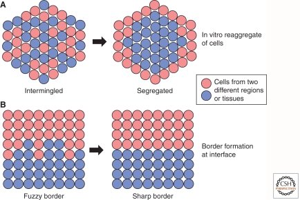
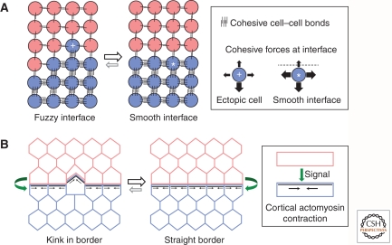
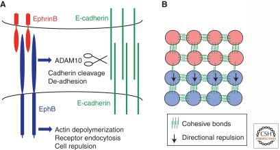
The establishment and maintenance of precisely organized tissues requires the formation of sharp borders between distinct cell populations. The maintenance of segregated cell populations is also required for tissue homeostasis in the adult, and deficiencies in segregation underlie the metastatic spreading of tumor cells. Three classes of mechanisms that underlie cell segregation and border formation have been uncovered. The first involves differences in cadherin-mediated cell-cell adhesion that establishes interfacial tension at the border between distinct cell populations. A second mechanism involves the induction of actomyosin-mediated contraction by intercellular signaling, such that cortical tension is generated at the border. Third, activation of Eph receptors and ephrins can lead to both decreased adhesion by triggering cleavage of E-cadherin, and to repulsion of cells by regulation of the actin cytoskeleton, thus preventing intermingling between cell populations. These mechanisms play crucial roles at distinct boundaries during development, and alterations in cadherin or Eph/ephrin expression have been implicated in tumor metastasis.


Figures

Figure 1.
Figure 1.
Cell segregation and boundary formation. (A) In vitro assay for cell segregation in which two cell populations are dissociated, mixed, and reaggregated. Over a period of time, the intermingled cell populations may segregate from each other. When performed with one population expressing lower levels of cadherin (red) than the other (blue), the former segregate to the outer region of the reaggregate, as predicted by the differential adhesion hypothesis. (B) During normal development, distinct tissues or regional domains within tissues are induced to form at specific locations. In many cases, the border of the adjacent domains is initially fuzzy, and local cell segregation underlies the formation of a sharp interface.
Figure 2.
Figure 2.
Cell adhesion and cortical tension. (A) One mechanism underlying cell segregation is mediated by differential adhesion of the two distinct cell populations. The differential adhesion hypothesis proposes that segregation is driven by the aggregate achieving the lowest free energy in which there is maximal contact between cells with the higher mutual affinity. At the interface of the two populations, interfacial tension occurs because the more adhesive cells (blue) will experience stronger cohesive forces from “like” cells than from “unlike” cells (red). The cohesive forces experienced by an ectopic cell (+) or by a cell at a smooth interface (*) are illustrated. At a fuzzy interface there is a net decrease in cohesive bonds and increase in free energy compared with a smooth interface. (B) Another mechanism that underlies boundary sharpening and maintenance is mediated by cortical tension. Signaling from one cell population (red) induces assembly and contraction of cell-surface-associated actomyosin in the adjacent cells (blue). This cortical tension sharpens the borders and restrains movement of cells across the interface.
Figure 3.
Figure 3.
Eph receptor and ephrin signaling. (A) Interactions between EphB receptor and ephrinB ligand can trigger two mechanisms that underlie cell segregation. The first is mediated by interactions of EphB receptor with the metalloproteinase ADAM10 and with E-cadherin. On binding of EphB receptor to ephrinB ligand, ADAM10 activity cleaves E-cadherin, thus leading to decreased adhesion selectively at the Eph-ephrin interface. The second mechanism involves signaling pathways downstream from activated EphB receptor that lead to depolymerization of the actin cytoskeleton, with concommitant endocytosis of Eph-ephrin complexes. The combination of cytoskeletal collapse and cell disengagement leads to repulsion of EphB- by ephrinB-expressing cells. (B) Decreased E-cadherin-mediated adhesion between EphB and ephrinB cells establishes differential adhesion. Directional repulsion of EphB-expressing cells by ephrinB-expressing cells prevents intermingling. These two mechanisms may act together to establish and maintain cell segregation.

References

    1. Abercrombie M 1979. Contact inhibition and malignancy. Nature 281: 259–262 - PubMed
    1. Abercrombie M, Heaysman JE 1953. Observations on the social behaviour of cells in tissue culture. I. Speed of movement of chick heart fibroblasts in relation to their mutual contacts. Exp Cell Res 5: 111–131 - PubMed
    1. Astin JW, Batson J, Kadir S, Charlet J, Persad RA, Gillatt D, Oxley JD, Nobes CD 2010. Competition amongst Eph receptors regulates contact inhibition of locomotion and invasiveness in prostate cancer cells. Nat Cell Biol 12: 1194–1204 - PubMed
    1. Barrios A, Poole RJ, Durbin L, Brennan C, Holder N, Wilson SW 2003. Eph/ephrin signaling regulates the mesenchymal-to-epithelial transition of the paraxial mesoderm during somite morphogenesis. Curr Biol 13: 1571–1582 - PubMed
    1. Batlle E, Henderson JT, Beghtel H, van den Born MM, Sancho E, Huls G, Meeldijk J, Robertson J, van de Wetering M, Pawson T, et al. 2002. β-catenin and TCF mediate cell positioning in the intestinal epithelium by controlling the expression of EphB/ephrinB. Cell 111: 251–263 - PubMed

Publication types

LinkOut - more resources