. 2012 Mar;56(3):1548-56.
doi: 10.1128/AAC.05100-11. Epub 2012 Jan 3.

Lactoferrin inhibits Porphyromonas gingivalis proteinases and has sustained biofilm inhibitory activity

Affiliations

Lactoferrin inhibits Porphyromonas gingivalis proteinases and has sustained biofilm inhibitory activity

Stuart G Dashper et al. Antimicrob Agents Chemother. 2012 Mar.

Abstract

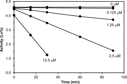
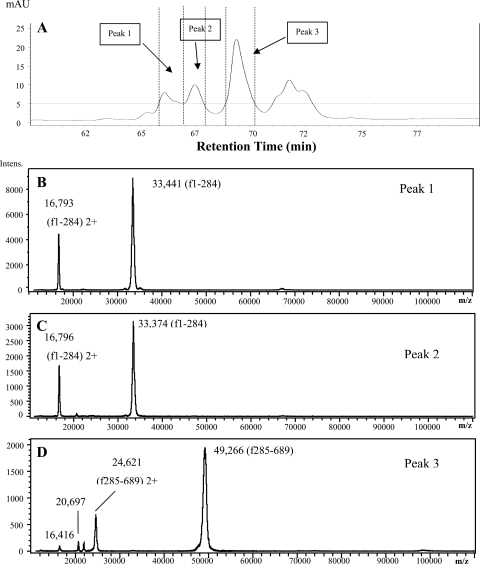
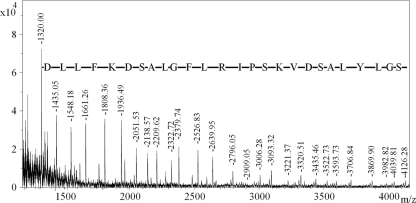
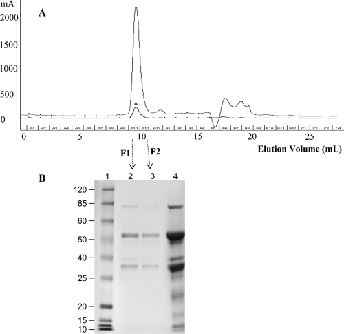
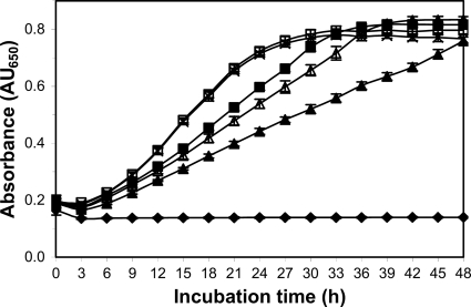
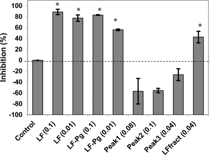
Porphyromonas gingivalis is a bacterial pathogen associated with chronic periodontitis that results in destruction of the tooth's supporting tissues. The major virulence determinants of P. gingivalis are its cell surface Arg- and Lys-specific cysteine proteinases, RgpA/B and Kgp. Lactoferrin (LF), an 80-kDa iron-binding glycoprotein found in saliva and gingival crevicular fluid, is believed to play an important role in innate immunity. In this study, bovine milk LF displayed proteinase inhibitory activity against P. gingivalis whole cells, significantly inhibiting both Arg- and Lys-specific proteolytic activities. LF inhibited the Arg-specific activity of purified RgpB, which lacks adhesin domains, and also inhibited the same activity of the RgpA/Kgp proteinase-adhesin complexes in a time-dependent manner, with a first-order inactivation rate constant (k(inact)) of 0.023 min(-1) and an inhibitor affinity constant (K(I)) of 5.02 μM. LF inhibited P. gingivalis biofilm formation by >80% at concentrations above 0.625 μM. LF was relatively resistant to hydrolysis by P. gingivalis cells but was cleaved into two major polypeptides (53 and 33 kDa) at R(284) to S(285), as determined by in-source decay mass spectrometry; however, these polypeptides remained associated with each other and retained inhibitory activity. The biofilm inhibitory activity of LF against P. gingivalis was not attributed to direct antibacterial activity, as LF displayed little growth inhibitory activity against planktonic cells. As the known RgpA/B and Kgp inhibitor N-α-p-tosyl-l-lysine chloromethylketone also inhibited P. gingivalis biofilm formation, the antibiofilm effect of LF may at least in part be attributable to its antiproteinase activity.


Figures

Fig 1
Fig 1
Time-dependent inhibition of RgpA activity by lactoferrin. The figure shows the effect of lactoferrin at different concentrations (μM) on the activity (Ln%) of RgpA over time. The slope of these lines provided the apparent inactivation rate constants (kapp). Plotting 1/kapp versus 1/[I], where I is the lactoferrin concentration, yielded a straight line (r = 0.9995) with an intercept of 1/kinact and a slope of KI/kinact (28). This produced a first-order inactivation rate constant (kinact) of 0.023 min−1 and a lactoferrin binding affinity (KI) of 5.02 μM.
Fig 2
Fig 2
An orthographic view of the modeled lactoferrin-RgpB complex. The solvent-accessible surface of RgpB is shown in green, and the dark blue, space-filling atom is the zinc ion bound to the catalytic histidine (His244) of RgpB. Lactoferrin is shown as a ribbon structure (β-strand in yellow, α-helix in magenta, and coil in cyan), and the side-chains of residues that moved to within 3 Å of RgpB during the dynamics simulation are shown as “capped sticks.” The location of the atoms of the RgpB inhibitor, DFFR-chloromethylketone, are shown by red space-filling atoms.
Fig 3
Fig 3
(A) SDS-PAGE of LF incubated with P. gingivalis. Lane 1, LF (1 μg); lanes 2 to 7, LF and P. gingivalis; lane 2, 1-min incubation; lane 3, 10-min incubation; lane 4, 3-h incubation; lane 5, 6-h incubation; lane 6, 18-h incubation; lane 7, 3-day incubation. The arrow indicates the sample fractionated by gel filtration (6-h incubation). Major fragments of LF are labeled with Roman numerals, and the gel bands have been analyzed by MS. (B) LF was treated with different concentrations of trypsin at 37°C for 18 h. Lane 1, LF/trypsin = 2,000/1; lane 2, LF/trypsin = 5,000/1.
Fig 4
Fig 4
The primary sequence of LF. Underlined sequences denote the peptides identified by peptide mass fingerprint (PMF) analysis of fragments I to V. The cleavage site (‖) between fragments I and IV (the major polypeptides in the LF-Pg sample) was determined by in-source decay (ISD) MALDI-TOF MS. All arginine and lysine residues are shown in bold.
Fig 5
Fig 5
(A) RP-HPLC analysis of LF after 6 h of hydrolysis by P. gingivalis whole cells. (B to D) MS spectra of RP-HPLC peaks 1 to 3, respectively.
Fig 6
Fig 6
ISD MS spectrum of fragment I of LF-Pg. The fragment ions labeled correspond to N-terminal (C-type) fragments of lactoferrin, starting from C-12 and extending to C-38. The mass difference between each peak corresponds to the amino acid residue shown. Extrapolation of these data indicated that the N-terminal sequence of this polypeptide was 285SFQLFGSPPGQRDLLFKDSALGFLRIPSKVDSALYLGS, with the underlined portion evident in the spectrum.
Fig 7
Fig 7
(A) LF-Pg was fractionated by size-exclusion chromatography (SEC), while native LF was eluted as a control (indicated by an asterisk). The fractions were collected at an interval of 1 ml, and two fractions (marked as F1 and F2) were collected from the peak at the elution volume between 9 and 11 ml. (B) LF-Pg and the SEC fractions from LF-Pg were analyzed by using SDS-PAGE. Lane 1, MW marker; lanes 2 and 3, F1 and F2 from the peak eluted from SEC; lane 4, LF-Pg without SEC.
Fig 8
Fig 8
Effect of LF on P. gingivalis biofilm formation. P. gingivalis biofilm formation was determined in the presence of LF for 18 h (△), 24 h (▲), and 48 h (■). Each data point represents the mean and standard deviation of three replicates. Note the categorical scale on the x axis.
Fig 9
Fig 9
Effect of LF on planktonic growth of P. gingivalis in batch culture, LF concentrations (mg/ml): 0 (×), 0.5 (□), 2.5 (■), 5.0 (△), 10 (▲), uninoculated growth medium (♦). Each data point represents the mean and standard deviation of six biological replicates. LF concentration at 10 mg/ml, 5.0 mg/ml, and 2.5 mg/ml had a significant (P < 0.05) but moderate lowering of the growth rate but did not significantly effect final absorbance of the cultures.
Fig 10
Fig 10
Comparison of the biofilm inhibitory activity of native LF, LF-Pg (6 h of incubation), and fractions obtained from RP-HPLC separation of LF-Pg. Each data point represents the mean of three biological replicates. Peaks 1 to 3 correspond to the three RP-HPLC fractions in Fig. 4. LFfract was native LF eluted under the same conditions as that for separation of LF-Pg sample. The number in each treatment represents the protein concentration at mg/ml. *, significantly (P < 0.001) inhibited biofilm formation.

Similar articles

Cited by

References

    1. Albandar JM, Brunelle JA, Kingman A. 1999. Destructive periodontal disease in adults 30 years of age and older in the United States, 1988-1994. J. Periodontol. 70:13–29 - PubMed
    1. Anderson BF, et al. 1987. Structure of human lactoferrin at 3.2-Å resolution. Proc. Natl. Acad. Sci. U. S. A. 84:1769–1773 - PMC - PubMed
    1. Armfield J, Roberts-Thomson K, Spencer A. 2000. Australia's health 2000, vol 7. Catalogue no. AUS 19. Australian Institute of Health and Welfare, Canberra, Australia
    1. Arnold RR, Russell JE, Champion WJ, Brewer M, Gauthier JJ. 1982. Bactericidal activity of human lactoferrin: differentiation from the stasis of iron deprivation. Infect. Immun. 35:792–799 - PMC - PubMed
    1. Bhogal PS, Slakeski N, Reynolds EC. 1997. A cell-associated protein complex of Porphyromonas gingivalis W50 composed of Arg- and Lys-specific cysteine proteinases and adhesins. Microbiology 143:2485–2495 - PubMed

Publication types

MeSH terms