. 2012 Jan 3:3:610.
doi: 10.1038/ncomms1607.

Structure-based mutagenesis reveals the albumin-binding site of the neonatal Fc receptor

Affiliations
Free PMC article

Structure-based mutagenesis reveals the albumin-binding site of the neonatal Fc receptor

Jan Terje Andersen et al. Nat Commun. .
Free PMC article

Abstract

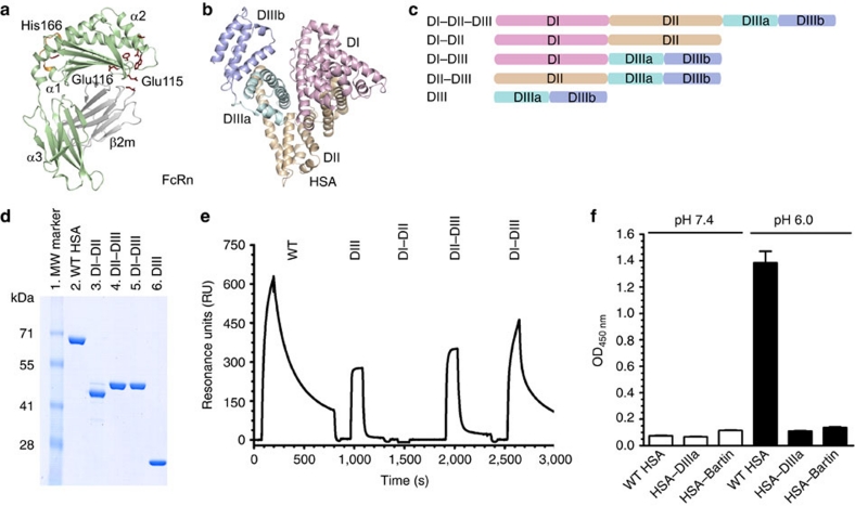
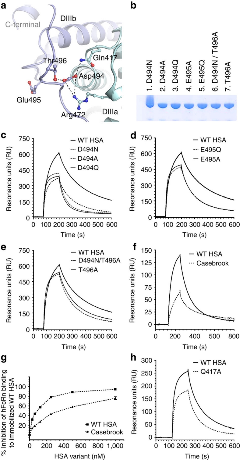
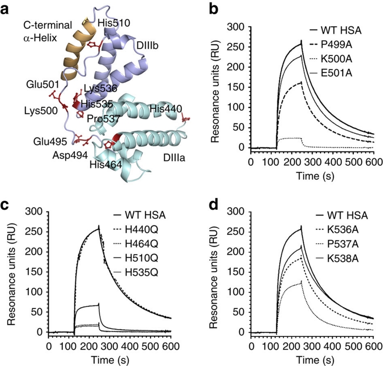
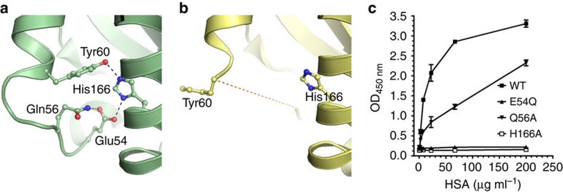
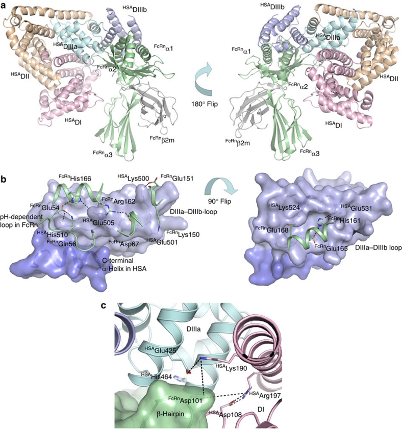
Albumin is the most abundant protein in blood where it has a pivotal role as a transporter of fatty acids and drugs. Like IgG, albumin has long serum half-life, protected from degradation by pH-dependent recycling mediated by interaction with the neonatal Fc receptor, FcRn. Although the FcRn interaction with IgG is well characterized at the atomic level, its interaction with albumin is not. Here we present structure-based modelling of the FcRn-albumin complex, supported by binding analysis of site-specific mutants, providing mechanistic evidence for the presence of pH-sensitive ionic networks at the interaction interface. These networks involve conserved histidines in both FcRn and albumin domain III. Histidines also contribute to intramolecular interactions that stabilize the otherwise flexible loops at both the interacting surfaces. Molecular details of the FcRn-albumin complex may guide the development of novel albumin variants with altered serum half-life as carriers of drugs.


Conflict of interest statement

I.S., J.T.A., B.D., J.C., A.P., L.E. and D.S. are co-inventors on pending patent applications, which are entitled 'Albumin Variants' and relate to the data described in this paper. The remaining authors have no competing financial interest to declare.

Figures

Figure 1
Figure 1. Domain architecture of HSA and hFcRn binding properties of HSA hybrid molecules.
(a) Overall structure of hFcRn showing the location of the pH-dependent flexible loop (orange) and His-166 relative to the IgG binding site (red residues in ball-and-stick). (b) The crystal structure of full-length HSA shows three α-helical domains; DI (pink), DII (orange) and DIII (cyan/blue). The DIII is split into subdomains DIIIa (cyan) and DIIIb (blue). (c) Domain organization of constructed hybrid HSA molecules (DI–DII, DI–DIII, DII-DIII, DIII). (d) SDS–PAGE gel migration of the HSA domain variants. (e) Representative SPR sensorgrams of equal amounts of WT HSA and domain combinations injected over immobilized hFcRn at pH 6.0. (f) ELISA showing pH-dependent binding of 5 μg ml−1 of WT HSA, HSA–DIIIa and HSA–Bartin to hFcRn at pH 7.4 and pH 6.0. n=4. All data are presented as mean±s.d.
Figure 2
Figure 2. The structural implications of HSA Casebrook on hFcRn binding.
(a) Close-up view of the interaction network around Asp-494 in HSA. Asp-494 is located in the loop connecting subdomain DIIIa (cyan) and DIIIb (blue). Asp-494 forms an ionic interaction with Arg-472 and a hydrogen-bond interaction with Gln-417, both of which are located in subdomain DIIIa. Asp-494 also forms a hydrogen bond with Thr-496, thus stabilizing the loop connecting DIIIa and DIIIb. (b) SDS–PAGE gel migration of the mutants D494N, D494A, D494Q, E495Q, E495A, T496A and D494N/T496A. (c) Representative SPR sensorgrams showing binding of immobilized hFcRn to 1 μM of WT HSA and recombinantly produced Casebrook (D494N), D494A and D494Q. (d) E495Q and E495A and (e) T496A and D494N/T496A at pH 6.0. (f) Representative SPR sensorgrams of immobilized hFcRn binding to 1 μM of WT HSA and Casebrook isolated from a heterozygote patient. (g) Competitive binding of WT HSA and Casebrook to hFcRn at pH 6.0. The receptor (100 nM) was injected in the presence of titrated amounts of WT or Casebrook HSA (0.015–1,000 nM) over immobilized HSA. (h) SPR sensorgrams showing binding of immobilized hFcRn to 1 μM of WT HSA and Q417A at pH 6.0.
Figure 3
Figure 3. Conserved histidines are fundamental for binding to hFcRn.
(a) Structural location of selected residues in DIII of HSA. Residues in the loop connecting the subdomains DIIIa and DIIIb selected for mutagenesis (Asp-494, Glu-495, Lys-500 and Glu-501) as well as extra residues close to the connecting loop, such as the conserved histidines (His-464, His-510 and His-536), Lys-536 and Pro-537 are displayed as ball-and-stick (maroon). The non-conserved His-440 is distally localized. The last C-terminal α-helix is highlighted in yellow. Representative SPR sensorgrams showing binding of immobilized hFcRn to 1 μM of WT HSA and (b) P499A, K500A and E501A, and (c) H440Q, H464Q, H510Q and H535Q as well as (d) K536A, P537A and K538A at acidic pH (6.0).
Figure 4
Figure 4. His-166 of hFcRn stabilizes a flexible loop in a pH-dependent manner.
Close-up view of the FcRn HC loop area at different pH conditions. (a) At low pH (4.2), the positively charged His-166 forms charge-stabilized hydrogen-bond interactions with Glu-54 and Tyr-60 within the surface-exposed loop in hFcRn. (b) At high pH (8.2), the uncharged His-166 looses the interactions with Glu-54 and Tyr-60, and the loop between residues Trp-51 and Tyr-60 becomes flexible and structurally disordered (represented by the dashed line). (c) Binding of 0.5 μg ml−1 of hFcRn WT and mutants (E54Q, Q56A and H166A) to titrated amount of HSA (0.3–200 μg ml−1) coated in ELISA wells at pH 6.0. n=4. All data are presented as mean±s.d.
Figure 5
Figure 5. A proposed hFcRn-HSA docking model.
(a) An overview of the docked molecules in two orientations showing the FcRn HC (green), β2m (grey) and the three HSA α-helical domains DI (pink), DII (orange) and DIII (cyan/blue). The HSA DIII is split into DIIIa (cyan) and DIIIb (blue). (b) Close-up view of the interaction interface between hFcRn (green cartoon) and HSA (blue surface) in the docking model. The C-terminal end of HSA (dark blue) and the loop corresponding to residues 490–510 between subdomains DIIIa and DIIIb form a crevice on the HSA surface into which the pH-dependent and flexible loop in hFcRn (residues 51–59) might bind. His-166 of hFcRn may form strong, charge-stabilized interactions with HSA residues Glu-54 and Glu-505. HSA Glu-505 could further interact with hFcRn Arg-162. Possible salt-bridges are formed between Lys-150 and Glu-151 of hFcRn with Glu-501 and Lys-500 of HSA. A cleft on the HSA surface is formed between the loop connecting DIIIa and DIIIb and the α-helix encompassing residues 520–535. His-161 of hFcRn may interact with Glu-531 of HSA at low pH, and the complex could be further reinforced by the salt-bridge between hFcRn Glu-168 and HSA Lys-524. (c) Interaction interface between hFcRn (green surface) and HSA (pink, blue and cyan cartoon) in the docking model. A β-hairpin loop in hFcRn is wedged in-between domains DI (pink) and DIIIa (cyan) in HSA. The hFcRn Asp-110 could be a partner to either Lys-190 or Arg-197 of HSA following some structural rearrangements in this interface. The conserved His-464 is located in the DIIIa α-helix contacting the β-hairpin loop.

References

    1. Peters T. Jr. Serum albumin. Adv. Protein Chem. 37, 161–245 (1985). - PubMed
    1. Chaudhury C. et al.. The major histocompatibility complex-related Fc receptor for IgG (FcRn) binds albumin and prolongs its lifespan. J. Exp. Med. 197, 315–322 (2003). - PMC - PubMed
    1. Anderson C. L. et al.. Perspective–FcRn transports albumin: relevance to immunology and medicine. Trends Immunol. 27, 343–348 (2006). - PubMed
    1. Roopenian D. C. & Akilesh S. FcRn: the neonatal Fc receptor comes of age. Nat. Rev. Immunol. 7, 715–725 (2007). - PubMed
    1. Ward E. S. & Ober R. J. Chapter 4: Multitasking by exploitation of intracellular transport functions the many faces of FcRn. Adv. Immunol. 103, 77–115 (2009). - PMC - PubMed

Publication types

MeSH terms