. 2012 May;221(2):285-95.
doi: 10.1007/s00213-011-2621-9. Epub 2012 Jan 4.

Cigarette smoking and white matter microstructure

Affiliations

Cigarette smoking and white matter microstructure

Matthew Hudkins et al. Psychopharmacology (Berl). 2012 May.

Abstract

Rationale: Diffusion tensor imaging has been used before in testing associations between cigarette smoking and white matter integrity, with inconsistent results. Published reports indicate higher fractional anisotropy (FA, a measure of linear water diffusion) in some brain regions and lower FA in others in adult smokers compared to nonsmokers. Adolescent smokers exhibited elevated FA at several brain regions and a positive correlation of FA in the genu corpus callosum with exposure to smoking (pack-years).

Objective: To help resolve prior discrepancies, we studied adults, sampling multiple brain regions, and testing for relationships to clinical features of nicotine dependence and exposure to smoking.

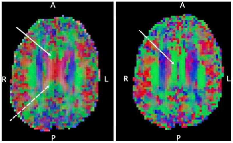
Methods: Brain MRI scans (1.5 T) were acquired, and FA and apparent diffusion coefficient (ADC, a measure of random diffusion) were assayed in corpus callosum and prefrontal white matter, corona radiata, internal capsule, cingulum bundle, and hippocampal perforant fibers in 18 smokers (33.7 ± 7.9 years of age) and 18 age- and gender-matched nonsmokers.

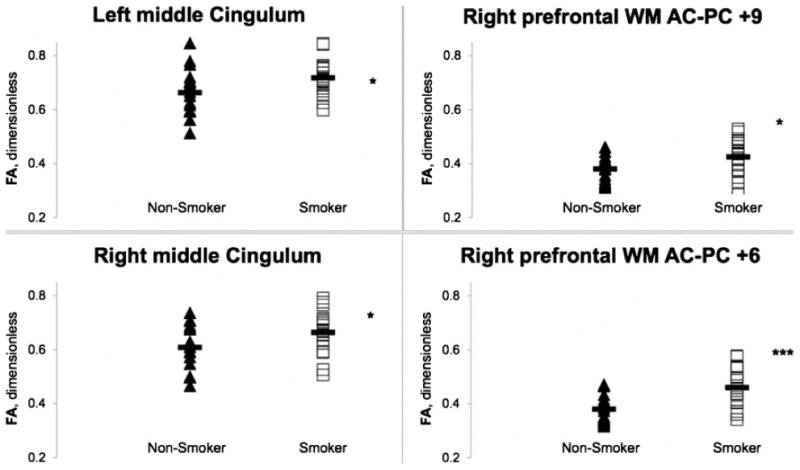
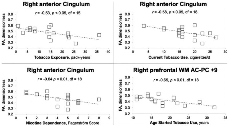
Results: ADC showed no group difference, but smokers had higher (4.3-21.1%) FA than nonsmokers. The differences were significant in right prefrontal white matter, cingulum, and genu corpus callosum. FA in several regions was negatively correlated with nicotine dependence or cigarettes/day.

Conclusions: Combined with earlier findings, these results suggest a model of changing trajectories whereby FA is higher with tobacco exposure during adolescence and declines with continued smoking in adulthood. This notion is supported by the observation that, at multiple sampling sites, participants who had started smoking earlier in life had higher FA than those who had started later.


Figures

Fig. 1
Fig. 1
Axial-oblique (parallel to AC-PC line) DTI color maps of the brain at level of (left) and above (right) the upper lateral ventricles from a selected participant. Red denotes fibers in the left-right direction, green denotes fibers in the anterior-posterior direction, blue denotes fibers in the inferior-superior direction. In the left panel, the DTI voxel selection of the anterior site of the cingulum white matter tract (solid white arrow) and posterior site of the cingulum white matter tract (dashed white arrow) are shown. These are the relatively small anterior-posterior oriented tracts mesial and anterior/posterior respectively to the corpus callosum as it forms a prominent “X” at the level of the upper lateral ventricles. In the right panel, the DTI voxel selection of the right middle cingulum site (dot-dashed white arrow) on a supraventricular axial-oblique slice is shown, centered in the midcaudally situated anterior-posterior tract just mesial to the superior corona radiata.
Fig. 2
Fig. 2
DTI FA whole-brain color map of data from a selected research participant. The image on the left shows an axial-oblique section (in the AC–PC plane). The white box in left prefrontal white matter (PFWM) indicates the boundaries of a 3-mm-deep, 9×9-mm2 slab, across which FA values were sampled and averaged in post-processing. A similar average was obtained for the right PFWM. The image on the right shows a parasagittal section of the same color map, depicting the positions of the four parallel prefrontal white-matter slabs (white bars) located at the level of the AC– PC plane (C), 6 mm below (D), and 6 (B) and 9 (A) mm above. Averages of left and right PFWM FA were obtained at each level.
Fig. 3
Fig. 3
Fractional anisotropy (FA) in nonsmokers (filled triangles) and smokers (open squares) at sites in left (upper left) and right (lower left) middle cingulum white-matter tracts. Data also shown (right) for FA averaged across each of two the 3-mm-thick, 9 × 9 mm2, axial-oblique, right prefrontal white-matter slabs. The slabs were oriented parallel to the AC-PC plane and centered 9 mm (upper right) and 6 mm (lower right) superior to it. Group mean FA (solid bar) was significantly higher for smokers than for nonsmokers in all four regions. Several other brain regions exhibited this elevation of FA in smokers (see Tables 2 and 3). p<*0.05, ***0.001 (protected post-hoc rank-transformed T-Test after omnibus repeated-measures MANCOVA covarying age and years of education).
Fig. 4
Fig. 4
Fractional anisotropy (FA) at sites in the right anterior cingulum bundle as a function of long-term tobacco exposure (pack-years, upper left), nicotine dependence (FTND score, lower left), and current tobacco use (cigarettes/day, upper right) in smokers. Data are also shown (lower right) for FA averaged across the 3-mm thick, 9 × 9 mm2, axial-oblique, right prefrontal white matter slab oriented parallel to the AC-PC plane and centered 9 mm superior to it, as a function of age when tobacco use began. Note significant negative correlations of FA with all three measures of smoking in same region of cingulum. Negative correlation of FA with FTND was observed at 2 other white matter sites; with current cigarette consumption at 2 other sites; and with long-term exposure at 2 other sites (Table 4). Protected post-hoc Spearman correlation following repeated-measures MANOVA for nicotine dependence, current cigarette consumption, and long-term smoking exposure, exploratory for age when smoking began.

Similar articles

Cited by

References

    1. Almeida OP, Garrido GJ, Lautenschlager NT, Hulse GK, Jamrozik K, Flicker L. Smoking is associated with reduced cortical regional gray matter density in brain regions associated with incipient Alzheimer disease. Am J Geriatr Psychiatry. 2008;16:92–98. - PubMed
    1. Azizian A, Monterosso J, O'Neill J, London ED. Magnetic resonance imaging studies of cigarette smoking. Handb Exp Pharmacol. 2009;192:113–143. - PubMed
    1. Bartzokis G. Acetylcholinesterase inhibitors may improve myelin integrity. Biol Psychiatry. 2007;62:294–301. - PubMed
    1. Bartzokis G, Lu PH. Brain Volume: Age-Related Changes. In: Squire LR, editor. Encyclopedia of Neuroscience. Vol. 2. Academic Press; Oxford: 2009. pp. 417–447.
    1. Bartzokis G, Lu PH, Stewart SB, Oluwadara B, Lucas AJ, Pantages J, et al. In vivo evidence of differential impact of typical and atypical antipsychotics on intracortical myelin in adults with schizophrenia. Schizophr Res. 2009;113:322–331. - PMC - PubMed

Publication types

LinkOut - more resources