. 2012 Jan;42(1):147-57.
doi: 10.1002/eji.201141927.

HIV-1 infection alters CD4+ memory T-cell phenotype at the site of disease in extrapulmonary tuberculosis

Affiliations
Free PMC article

HIV-1 infection alters CD4+ memory T-cell phenotype at the site of disease in extrapulmonary tuberculosis

Kerryn Matthews et al. Eur J Immunol. 2012 Jan.
Free PMC article

Abstract

HIV-1-infected people have an increased risk of developing extrapulmonary tuberculosis (TB), the immunopathogenesis of which is poorly understood. Here, we conducted a detailed immunological analysis of human pericardial TB, to determine the effect of HIV-1 co-infection on the phenotype of Mycobacterium tuberculosis (MTB)-specific memory T cells and the role of polyfunctional T cells at the disease site, using cells from pericardial fluid and blood of 74 patients with (n = 50) and without (n = 24) HIV-1 co-infection. The MTB antigen-induced IFN-γ response was elevated at the disease site, irrespective of HIV-1 status or antigenic stimulant. However, the IFN-γ ELISpot showed no clear evidence of increased numbers of antigen-specific cells at the disease site except for ESAT-6 in HIV-1 uninfected individuals (p = 0.009). Flow cytometric analysis showed that CD4+ memory T cells in the pericardial fluid of HIV-1-infected patients were of a less differentiated phenotype, with the presence of polyfunctional CD4+ T cells expressing TNF, IL-2 and IFN-γ. These results indicate that HIV-1 infection results in altered phenotype and function of MTB-specific CD4+ T cells at the disease site, which may contribute to the increased risk of developing TB at all stages of HIV-1 infection.


Figures

Figure 1
Figure 1
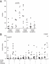
Pericardial TB patients have increased antigen-specific IFN-γ secretion in the pericardial fluid compared with blood. Undiluted blood and pericardial fluid were cultured overnight either unstimulated (NIL) or stimulated with ESAT-6 and CFP-10. IFN-γ concentration (ng/mL) was determined by ELISA and bars represent medians. Each circle is representative of one sample. P-Values indicate the differences between blood and pericardial fluid using the Wilcoxon-matched pairs test.
Figure 2
Figure 2
Increased numbers of ESAT-6-specific IFN-γ-producing cells in the pericardial fluid of HIV-1-uninfected patients. PBMCs and PFCs were assessed for antigen-specific IFN-γ responses by ELISpot assay. Each circle represents the number of IFN-γ spot forming cells/106 cells in response to ESAT-6 and CFP-10, corrected for unstimulated control wells. The number of ESAT-6 specific IFN-γ spot forming cells/106 cells was significantly higher in the pericardial fluid of the HIV-1-uninfected patients compared with HIV-1-infected patients (p=0.009, Mann–Whitney U test).
Figure 3
Figure 3
Flow cytometric analysis of CD4+ memory T cells in the pericardial fluid. (A) PFCs from HIV-1-uninfected (n=8, closed circles) and HIV-1-infected (n=7, open circles) patients were stimulated overnight in the presence of hkH37Rv, followed by surface staining for four-colour flow cytometry using CD4+, CD45RA and the co-stimulatory marker CD28. A significantly higher proportion of CD28+CD45RA cells at the disease site of the HIV-1-infected patients (p=0.04, Mann–Whitney U test), indicates a shift towards a less differentiated memory phenotype. Bars represent medians. (B) PFCs from HIV-1-uninfected (n=8, closed circles) and HIV-1-infected (n=10, open circles) patients were stimulated overnight in the presence of hkH37Rv, followed by surface staining for eight colour flow cytometry, including CD3, CD4, CD45RA, the co-stimulatory marker CD27 as well as CCR5 to phenotype the cells at the site of disease. The data indicate that CD4+ memory T cells in the pericardial fluid of HIV-1-infected patients lack the CCR5 receptor. A significantly higher proportion of CCR5CD27CD45RA cells (p=0.02), and significantly fewer CCR5+CD27+CD45RA+ cells (p=0.012) were found at the disease site of HIV-1-infected patients, compared with HIV-1-uninfected patients (Mann–Whitney U test). Bars represent medians.
Figure 4
Figure 4
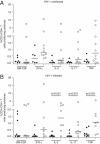
Increased cytokine-secreting capacity in the pericardium compared with that in blood, irrespective of HIV-1 status. Total cytokine secretion in response to ESAT-6+CFP-10 in blood (closed circles) and pericardial fluid (open circles) of (A) HIV-1-uninfected and (B) HIV-1-infected patients. Bars represent medians. Statistically significant differences between blood and fluid are indicated (Mann–Whitney U test).
Figure 5
Figure 5
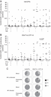
Polyfunctional CD4+ T cells are present at the disease site. CD3+CD4+ T cells were assessed for the expression of various combinations of IFN-γ, IL-2 and TNF in response to (A) hkH37Rv and (B) ESAT-6+CFP-10 stimulation, in the pericardial fluid of HIV-1-uninfected (closed circles) and HIV-1-infected (open circles) patients. (C) Pie-charts illustrate the overall proportional contribution of cytokine expressing cells to the antigen-specific response in blood and pericardial fluid. 1+: any one cytokine, 2+: any two cytokines, 3+: all three cytokines. IL-2 single-positive cells were proportionally expanded in the HIV-1-infected pericardial fluid compared with the uninfected in response to ESAT-6+CFP-10 (B, p=0.024, Mann–Whitney U test).
Figure 6
Figure 6
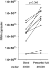
HIV viral load is increased in pericardial fluid compared with blood. HIV-1 RNA copies/mL were significantly elevated in matched pericardial fluid compared with serum from HIV-1-infected patients (p=0.002, Wilcoxon-matched pairs test). Squares represent the patient on ARV treatment at enrolment into the study.

Similar articles

Cited by

References

    1. Meintjes G, Wilkinson RJ. Undiagnosed active tuberculosis in HIV-infected patients commencing antiretroviral therapy. Clin. Infect. Dis. 2010;51:830–832. - PubMed
    1. WHO. Global tuberculosis control: key findings from the December 2009 WHO report. Wkly Epidemiol. Rec. 2010;85:69–80. - PubMed
    1. Wilkinson RJ, Vordermeier HM, Wilkinson KA, Sjolund A, Moreno C, Pasvol G, Ivanyi J. Peptide-specific T cell response to Mycobacterium tuberculosis: clinical spectrum, compartmentalization, and effect of chemotherapy. J. Infect. Dis. 1998;178:760–768. - PubMed
    1. Pathan AA, Wilkinson KA, Klenerman P, McShane H, Davidson RN, Pasvol G, Hill AV, et al. Direct ex vivo analysis of antigen-specific IFN-gamma-secreting CD4 T cells in Mycobacterium tuberculosis-infected individuals: associations with clinical disease state and effect of treatment. J. Immunol. 2001;167:5217–5225. - PubMed
    1. Mayosi BM, Wiysonge CS, Ntsekhe M, Gumedze F, Volmink JA, Maartens G, Aje A, et al. Mortality in patients treated for tuberculous pericarditis in sub-Saharan Africa. South Afr. Med. J. 2008;98:36–40. - PubMed

Publication types

MeSH terms