

Differentiation of rodent immune and hematopoietic system reactive lesions from neoplasias
- PMID: 22215512
- PMCID: PMC3443630
- DOI: 10.1177/0192623311431467
Differentiation of rodent immune and hematopoietic system reactive lesions from neoplasias
Abstract
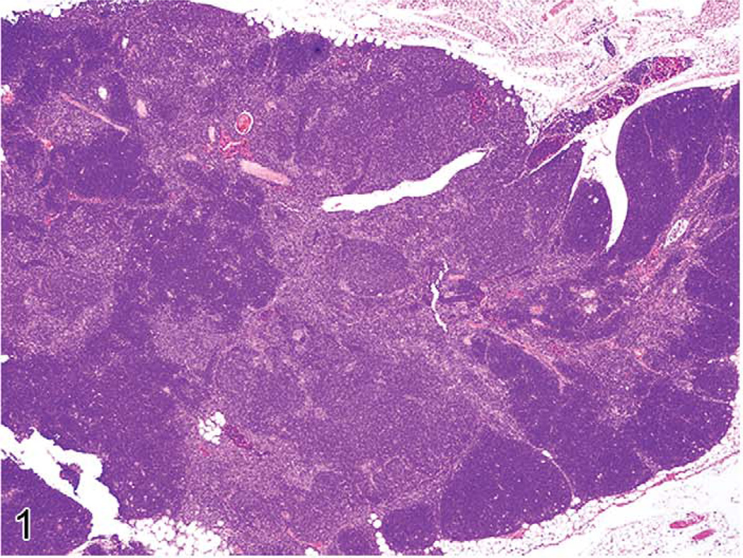
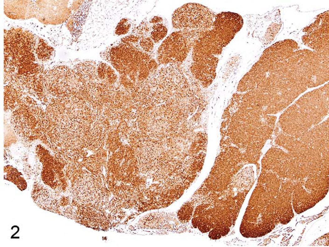
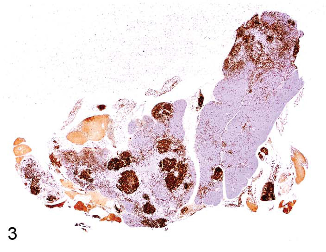
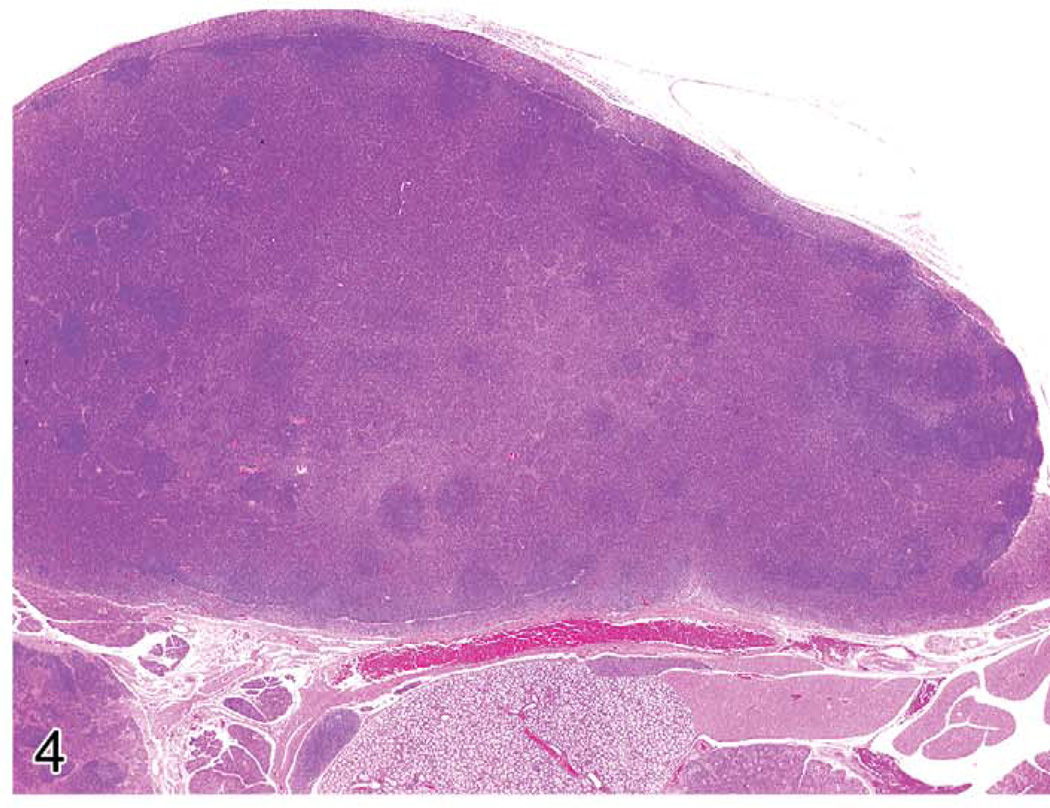
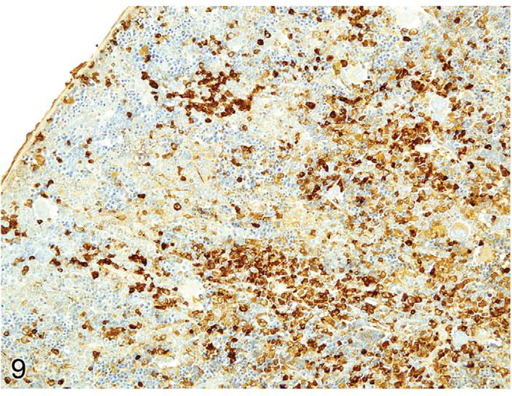
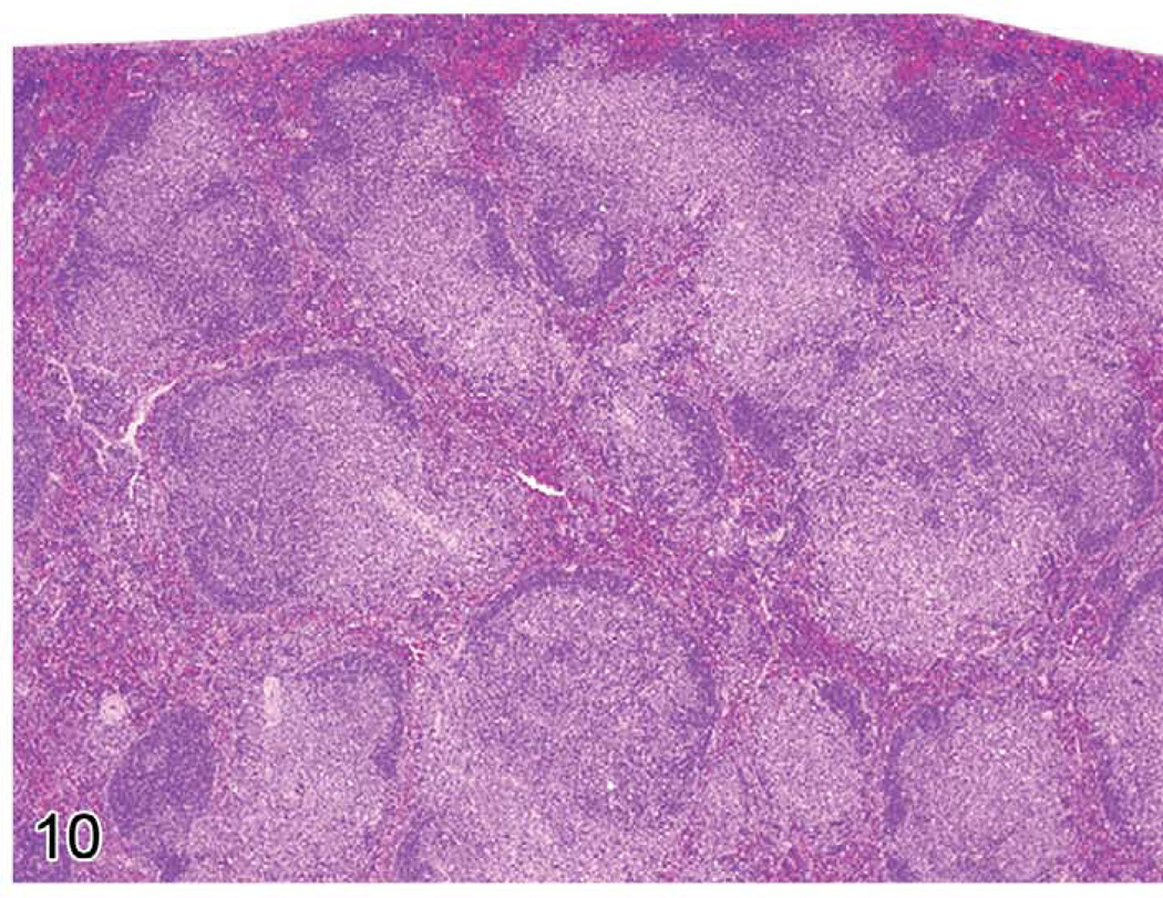
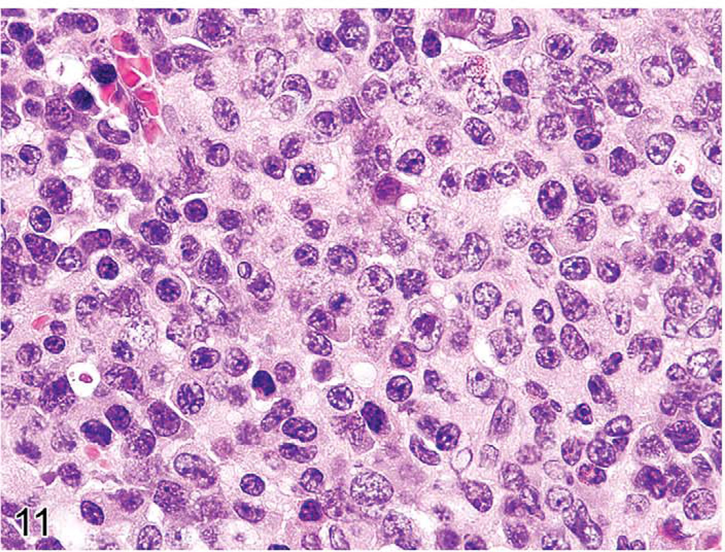
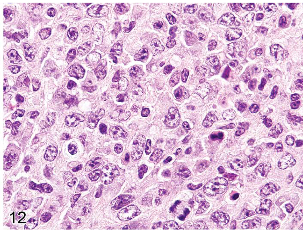
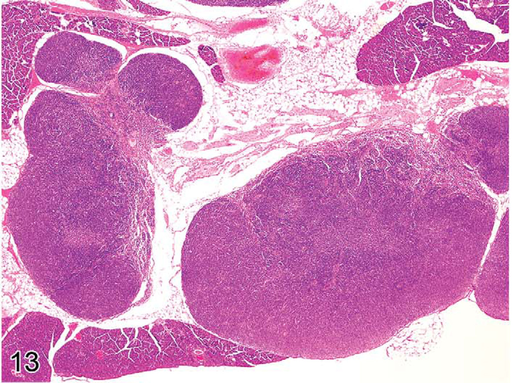
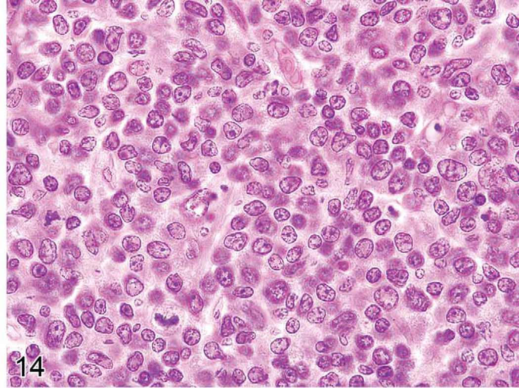
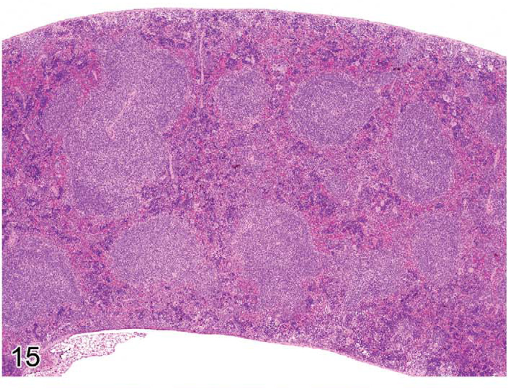
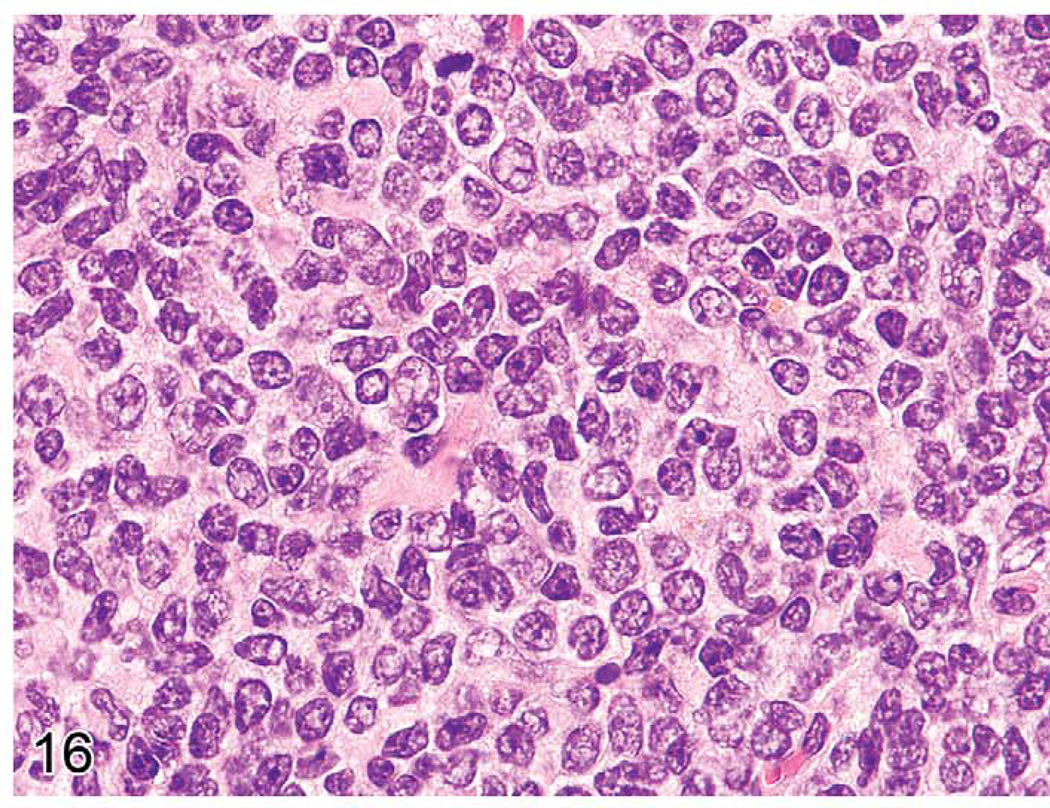
The immune and hematopoietic systems play an important role in the normal homeostasis of blood and blood cells and for immune responses to endogenous and exogenous processes and insults. In order to interpret histopathologic changes in the immune and hematopoietic systems, it is important to understand the normal anatomy and histology of the thymus, spleen, lymph nodes, bone marrow, and other tissues. The thymus, spleen, and lymph nodes can be categorized by anatomical compartments, each of which contributes to specific immune functions. Lesions may be diagnosed by interpretive or descriptive (semiquantitative) methods. The interpretation of these tissues by lesion in anatomical compartments should allow for better understanding of these reactions and more definitive pathologic findings. Proliferative lesions may be difficult to differentiate from lymphomas and leukemias. The use of immunohistochemistry, compartmental pathology, and methods for the evaluation of clonality will make interpretation easier.
Figures
Similar articles
-
Alpha4 integrins regulate the proliferation/differentiation balance of multilineage hematopoietic progenitors in vivo.Immunity. 1999 Nov;11(5):555-66. doi: 10.1016/s1074-7613(00)80131-4. Immunity. 1999. PMID: 10591181
-
The distribution of CD10 (NEP 24.11, CALLA) in humans and mice is similar in non-lymphoid organs but differs within the hematopoietic system: absence on murine T and B lymphoid progenitors.Eur J Immunol. 1995 Mar;25(3):677-87. doi: 10.1002/eji.1830250308. Eur J Immunol. 1995. PMID: 7705396
-
Pathology of acquired immunodeficiency syndrome (AIDS) in children.Keio J Med. 1996 Dec;45(4):306-12. doi: 10.2302/kjm.45.306. Keio J Med. 1996. PMID: 9023448 Review.
-
The utility of immunohistochemistry for the identification of hematopoietic and lymphoid cells in normal tissues and interpretation of proliferative and inflammatory lesions of mice and rats.Toxicol Pathol. 2012;40(2):345-74. doi: 10.1177/0192623311430695. Toxicol Pathol. 2012. PMID: 22434870 Review.
-
Reactive lymphoid hyperplasia of the liver.J Clin Gastroenterol. 1993 Apr;16(3):240-4. doi: 10.1097/00004836-199304000-00017. J Clin Gastroenterol. 1993. PMID: 8505499
Cited by
-
Correlation of in vivo imaging to morphomolecular pathology in translational research: challenge accepted.EJNMMI Res. 2021 Aug 28;11(1):83. doi: 10.1186/s13550-021-00826-2. EJNMMI Res. 2021. PMID: 34453623 Free PMC article.
-
IL-21-driven neoplasms in SJL mice mimic some key features of human angioimmunoblastic T-cell lymphoma.Am J Pathol. 2015 Nov;185(11):3102-14. doi: 10.1016/j.ajpath.2015.07.021. Epub 2015 Sep 9. Am J Pathol. 2015. PMID: 26363366 Free PMC article.
-
Protective effects of dietary grape against atopic dermatitis-like skin lesions in NC/NgaTndCrlj mice.Front Immunol. 2023 Jan 19;13:1051472. doi: 10.3389/fimmu.2022.1051472. eCollection 2022. Front Immunol. 2023. PMID: 36741360 Free PMC article.
-
Nanoparticle-mediated convection-enhanced delivery of a DNA intercalator to gliomas circumvents temozolomide resistance.Nat Biomed Eng. 2021 Sep;5(9):1048-1058. doi: 10.1038/s41551-021-00728-7. Epub 2021 May 27. Nat Biomed Eng. 2021. PMID: 34045730 Free PMC article.
-
Hematopoietic neoplasms in Prkar2a-deficient mice.J Exp Clin Cancer Res. 2015 Nov 25;34:143. doi: 10.1186/s13046-015-0257-z. J Exp Clin Cancer Res. 2015. PMID: 26608815 Free PMC article.
References
-
- Adams JM, Harris AW, Pinkert CA, Corcoran LM, Alexander WS, Cory S, Palmiter RD, Brinster RL. The c-myc oncogene driven by immunoglobulin enhancers induces lymphoid malignancy in transgenic mice. Nature. 1985;318:533–538. - PubMed
-
- Barton E, Mandal P, Speck SH. Pathogenesis and host control of gammaherpesviruses: Lessons from the mouse. Ann Rev Immunology. 2011;29:351–397. - PubMed
-
- Cattoretti G, Pasqualucci L, Ballon G, Tam W, Nandula SV, Shen Q, Mo T, Murty VV, Dalla-Favera R. Deregulated BCL6 expression recapitulates the pathogenesis of human diffuse large B cell lymphomas in mice. Cancer Cell. 2005;7:445–455. - PubMed
-
- Cesta MF. Normal structure, function, and histology of the spleen. Toxicol Pathol. 2006;34:455–465. - PubMed
Publication types
MeSH terms
Grants and funding
LinkOut - more resources
Full Text Sources
