Review
. 2012 Mar-Apr;3(2):195-212.
doi: 10.1002/wrna.1105. Epub 2012 Jan 3.

The structures of nonprotein-coding RNAs that drive internal ribosome entry site function

Affiliations
Review

The structures of nonprotein-coding RNAs that drive internal ribosome entry site function

Terra-Dawn M Plank et al. Wiley Interdiscip Rev RNA. 2012 Mar-Apr.

Abstract

Internal ribosome entry sites (IRESs) are RNA sequences that can recruit the translation machinery independent of the 5' end of the messenger RNA. IRESs are found in both viral and cellular RNAs and are important for regulating gene expression. There is great diversity in the mechanisms used by IRESs to recruit the ribosome and this is reflected in a variety of RNA sequences that function as IRESs. The ability of an RNA sequence to function as an IRES is conferred by structures operating at multiple levels from primary sequence through higher-order three-dimensional structures within dynamic ribonucleoproteins (RNPs). When these diverse structures are compared, some trends are apparent, but overall it is not possible to find universal rules to describe IRES structure and mechanism. Clearly, many different sequences and structures have evolved to perform the function of recruiting, positioning, and activating a ribosome without using the canonical cap-dependent mechanism. However, as our understanding of the specific sequences, structures, and mechanisms behind IRES function improves, more common features may emerge to link these diverse RNAs.


Figures

Figure 1
Figure 1
Comparison of the mechanism of cap-dependent and cap-independent translation initiation in eukaryotes. (a) In cap-dependent translation initiation, the m7G cap (cap) is bound by eIF4E (red) and this leads to the binding of additional initiation factors. Recruitment of the 40S ribosomal subunit (yellow) and associated factors occurs through the interaction of eIF3 (orange). The 40S subunit-containing preinitiation complex then scans to the start codon. Start codon recognition and GTP hydrolysis allows the initiation factors to dissociate and the 60S ribosomal subunit joins to create a translationally competent 80S ribosome. (b) In cap-independent internal translation initiation, RNA sequences called internal ribosome entry sites (IRESs) recruit the 40S subunit independently of the cap. The mechanism behind 40S recruitment varies between different IRES RNAs and may or may not require the use of additional initiation factors (dashed shapes). Depending on the IRES RNA, the 40S is either recruited directly or scans to the start codon. Once the start codon is recognized, the initiation factors dissociate (if used) and the 60S ribosomal subunit joins.
Figure 2
Figure 2
Examples of the diversity of mechanisms for ribosome recruitment by viral IRES RNAs. Viral IRESs for which mechanistic information is available has allowed them to be placed in several classes. In the Dicistroviridae IGR IRESs, the 40S subunit is recruited directly to the initiator codon, without the need for additional initiation factors or tRNA. Although not depicted in this figure, one report has shown that the IGR IRESs can bind directly to pre-formed 80S ribosomes. In other IRESs (HCV and similar) the 40S subunit directly binds the IRES but requires additional factors (eIF3, 2) for function. In still other IRESs (FMDV, EMCV), additional initiation factors are required for recruitment of the 40S subunit directly to the initiator codon. Finally, another set of IRESs (Poliovirus, HAV), not only require additional initiation factors to recruit the 40S subunit to the RNA but utilize a scanning mechanism for identification of the AUG start codon. A simplified pathway is shown, depicting how these different pathways compare. A possible relationship between the need for factors and the ability of an IRES RNA to form a stable 3-D fold is discussed elsewhere. Note that for many other IRESs, their mechanisms are unknown and thus they cannot yet be placed in a class. It also remains possible that some will have mechanisms different from anyxia of these.
Figure 3
Figure 3
How IRESs are identified and functionally studied and the caveats and considerations in these experiments. (a) IRESs typically are identified by placing the sequence of interest in front of a reporter construct and expression of the reporter is compared to a non-IRES control construct. Examples of the types of constructs used in these studies, the conditions under which they can be used, and the expected results of expression studies are depicted. For example, dual-cistronic reporter constructs can be used in both RNA and DNA transfections of cells, as well as in-vitro transcribed and programmed into lysate. The upstream reporter serves as an internal control for transfection efficiency and unlike their monocistronic counterparts, these constructs do not require a transfection control. Under normal eIF4E function, both reporters should be expressed if the insert contains an IRES. However, if the insert does not contain an IRES (such as in the non-IRES control construct), the downstream reporter should not be expressed. In contrast, under conditions where eIF4E is inhibited, the upstream reporter should not be expressed in either construct, while the downstream reporter will be expressed if the insert contains an IRES. (b) Transfection of dual-cistronic DNA constructs into cells can be subjected to nuclear processing events (dashed lines). These events include cryptic splicing (magenta) and cryptic promoter activity (green). If present, these result in the production of capped mono-cistronic RNA species (shown as the resultant RNAs) and can lead to false positive IRES reporter levels. (c) The use of dual-cistronic DNA constructs also can lead to ribosomal readthrough on the message (right) rather than correct termination and re-entry on the downstream reporter (left). This would yield false positive signal of IRES activity. (d) Capped, mono-cistronic constructs may recruit ribosomes through both a cap-dependent and cap-independent process, confounding clear delineation of the level of IRES activity. In this case, IRES identification must occur under conditions where cap-dependent translation is inhibited. (e) Diagram of a circular RNA containing an IRES upstream of a reporter sequence. Assuming the RNA remains intact during the experiment (e.g. – is not nicked), ribosomes can only enter in a cap- and end-independent mechanism; hence, expression of the reporter indicates an IRES.
Figure 4
Figure 4
Model for the mechanism of internal ribosome recruitment by the YMR181c IRES. In cap-dependent translation initiation, PABP binds to the poly-A tail at the 3′ end of the mRNA as well as to eIF4G at the 5′ end of the message (top). This 5′-3′ crosstalk leads to circularization of the message and efficient recycling of ribosomes. In contrast, PABP binds an A-rich sequence the 5′UTR of YMR181c, leading to recruitment of eIF4G and the downstream translation machinery (bottom). Thus, in YMR181c, binding of the PABP to the A-rich primary structure upstream of the AUG leads to recruitment of eIF4G independently of the cap and 4E interaction.
Figure 5
Figure 5
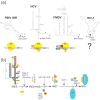
Examples in the diversity of IRES RNA secondary structures and the role of secondary structure in mechanism. (a) Secondary structure models of several viral IRESs and diagrams of their factor requirements for 40S recruitment. In the case of HIV-1 5′UTR IRES, the secondary structure is known,, while the mechanism behind internal 40S recruitment is still unknown. (b) Detailed diagram of the mechanism for RNA structure-based ribosome recruitment in the HCV-like IRESs. HCV-like IRESs bind the 40S subunit (yellow circle) directly using the base of domain III (boxed in yellow on the secondary structure diagram). Domain IIIb (orange box) recruits eIF2, leading to tRNA and eIF3 (green and orange circles) binding. The resulting complex is called the 48S*, asterisk denoting the deffierence between it and canonically assembled 48S. Domain II (blue box) triggers GTP hydrolysis and 60S recruitment (blue oval).
Figure 6
Figure 6
Further examples in the diversity of IRES RNA secondary structures. (a) Diagram of the Dicistroviridae genome, with the 5′UTR and intergenic region (IGR) IRESs highlighted (red and blue dashed boxes, respectively). Secondary structure diagrams of PSIV IRESs are shown. A proposed pseudoknot interaction is indicated with a line on the 5′ IRES diagram. (b) Secondary structure diagrams of the HCV, c-myc and PSIV IGR IRESs with pseudoknot structures depicted (shaded in red).
Figure 7
Figure 7
Cryo-EM reconstructions of the CrPV IGR IRES (top) and HCV IRES (bottom) bound to human 80S ribosomes. The 60S subunit is in cyan, the IRES RNA in purple and 40S subunit in yellow. Two views are shown for each, with the secondary structure cartoon of each IRES to the left. The overall differences in the global architectures of these two IRESs is obvious, as is the difference in their locations on the ribosome.
Figure 8
Figure 8
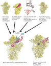
Example of how multiple sets of structural data can lead to a model. In this example, an x-ray crystal structure of the 40S subunit from Tetrahymena thermophila (top left, eIF1A is not shown), a cryo-EM structure of the CrPV IGR IRES bound to the ribosome (second from left, the 60S subunit is not shown), x-ray crystal structures of unbound IGR IRES domains (second from right, with secondary structure cartoon,, and the x-ray crystal structure of an IGR IRES domain bound to a 70S ribosome (top right, 50S subunit not shown) were combined to yield the model at lower left. In this model, the IGR IRES is shown in magenta, and two proteins known to interact with the IGR IRES are shown in cyan (rpS25) and green (rpS5). To the right of the IRES structure is the crystal structure of a 70S ribosome with bound tRNAs (50S subunit not shown). Note how the IGR IRES spans the binding sites of all three tRNAs.

Similar articles

Cited by

References

    1. Mathews MB, Sonenberg N, Hershey BJW. Origins and Principles of Translational Control. In: Mathews MB, Sonenberg N, Hershey JWB, editors. Translational Control in Biology and Medicine. Cold Spring Harbor, NY: Cold Spring Harbor Laboratory Press; 2007. pp. 1–40.
    1. Kozak M. An analysis of vertebrate mRNA sequences: Intimations of translational control. Journal of Cell Biology. 1991;115:887–903. - PMC - PubMed
    1. Kozak M. Point mutations define a sequence flanking the AUG initiator codon that modulates translation by eukaryotic ribosomes. Cell. 1986;44:283–292. - PubMed
    1. Jackson RJ, Hellen CU, Pestova TV. The mechanism of eukaryotic translation initiation and principles of its regulation. Nat Rev Mol Cell Biol. 2010;11:113–127. - PMC - PubMed
    1. Pestova T, Lorsch JR, Hellen CU. The Mechanism of Translation Initiation in Eukaryotes. In: Mathews MB, Sonenberg N, Hershey JWB, editors. Translational Control in Biology and Medicine. Cold Spring Harbor, NY: Cold Spring Harbor Laboratory Press; 2007. pp. 87–128.

Publication types

Substances

LinkOut - more resources