

doi: 10.1007/978-1-61779-504-6_5.
Measurement of mitochondrial oxygen consumption using a Clark electrode
Affiliations
- PMID: 22215541
- PMCID: PMC8711122
- DOI: 10.1007/978-1-61779-504-6_5
Item in Clipboard
Measurement of mitochondrial oxygen consumption using a Clark electrode
Methods Mol Biol.
2012.
Abstract
Mitochondria require oxygen to produce ATP in sufficient quantities to drive energy-requiring reactions in eukaryotic organisms. The measurement of oxygen consumption rates from isolated mitochondria in vitro is a useful and valuable technique in the research and evaluation of mitochondrial dysfunction and disease since ADP-dependent oxygen consumption directly reflects coupled respiration or oxidative phosphorylation (OXPHOS). This chapter describes the traditional method of mitochondrial polarography using a Clark electrode for measuring coupled respiration in freshly isolated mitochondria from both mammalian tissues and Drosophila melanogaster.
Figures
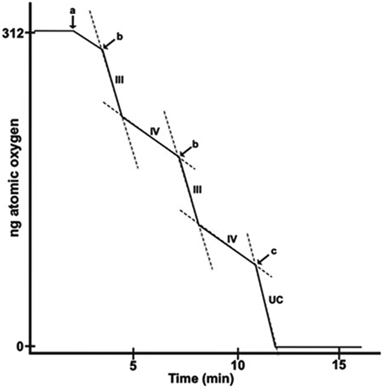
Idealized trace of a polarographic run. At time “0,” substrate (5 μM of glutamate + malate or succinate) is added to chamber full of air-saturated respiration buffer, and a stable baseline is observed. (a) Isolated mitochondria are added to a final concentration of 0.3 mg/mL. (b) 125 nmol of ADP is added, stimulating state III rate which transitions to state IV once exogenous ADP is consumed by OXPHOS. (c) DNP (50 μM) is added to stimulate maximal uncoupled rate (UC).
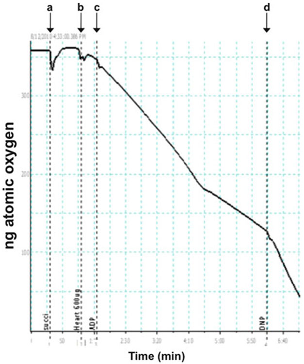
Typical polarographic trace of rat heart mitochondria with succinate as substrate. A polarographic experiment using mitochondria isolated from wild-type rat heart is shown. (a) Succinate (5 μM) is added. (b) Rat heart mitochondria (0.9 mg/mL) is added. (c) 125 nmol of ADP is added. (d) DNP (50 μM) is added.
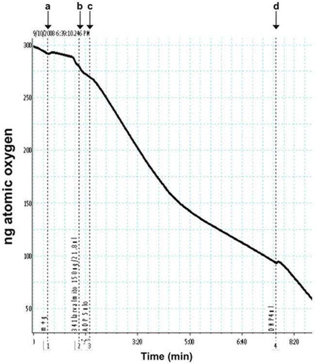
Typical polarographic trace of Drosophila larval mitochondria with glutamate + malate as substrate. A polarographic experiment using mitochondria isolated from wild-type Drosophila melanogaster third instar larvae is shown. (a) Glutamate + malate (5 μM each) is added. (b) Fly larval mitochondria (0.3 mg/mL) is added. (c) 125 nmol of ADP is added. (d) DNP (50 μM) is added.
Similar articles
-
Assessing Mitochondrial Bioenergetics by Respirometry in Cells or Isolated Organelles.Methods Mol Biol. 2018;1732:273-287. doi: 10.1007/978-1-4939-7598-3_18. Methods Mol Biol. 2018. PMID: 29480482
-
Evaluation of Respiration with Clark-Type Electrode in Isolated Mitochondria and Permeabilized Animal Cells.Methods Mol Biol. 2018;1782:7-29. doi: 10.1007/978-1-4939-7831-1_2. Methods Mol Biol. 2018. PMID: 29850992
-
Evaluation of Respiration with a Clark-Type Electrode in Isolated Mitochondria, Intact and Permeabilized Cells, and Explants from Animal Tissues.Methods Mol Biol. 2025;2878:1-34. doi: 10.1007/978-1-0716-4264-1_1. Methods Mol Biol. 2025. PMID: 39546254
-
In vivo and in organello assessment of OXPHOS activities.Methods. 2002 Apr;26(4):307-16. doi: 10.1016/S1046-2023(02)00036-1. Methods. 2002. PMID: 12054921 Review.
-
Mitochondrial Neurodegeneration: Lessons from Drosophila melanogaster Models.Biomolecules. 2023 Feb 16;13(2):378. doi: 10.3390/biom13020378. Biomolecules. 2023. PMID: 36830747 Free PMC article. Review.
Cited by
-
Macromitophagy is a longevity assurance process that in chronologically aging yeast limited in calorie supply sustains functional mitochondria and maintains cellular lipid homeostasis.Aging (Albany NY). 2013 Apr;5(4):234-69. doi: 10.18632/aging.100547. Aging (Albany NY). 2013. PMID: 23553280 Free PMC article.
-
Mitochondria as the Target of Hepatotoxicity and Drug-Induced Liver Injury: Molecular Mechanisms and Detection Methods.Int J Mol Sci. 2022 Mar 18;23(6):3315. doi: 10.3390/ijms23063315. Int J Mol Sci. 2022. PMID: 35328737 Free PMC article. Review.
-
Measurement of Oxygen Consumption Rate (OCR) and Extracellular Acidification Rate (ECAR) in Culture Cells for Assessment of the Energy Metabolism.Bio Protoc. 2018 May 20;8(10):e2850. doi: 10.21769/BioProtoc.2850. eCollection 2018 May 20. Bio Protoc. 2018. PMID: 34285967 Free PMC article.
-
Effects of arginine on coenzyme-Q10 micelle uptake for mitochondria-targeted nanotherapy in phenylketonuria.Drug Deliv Transl Res. 2024 Jan;14(1):191-207. doi: 10.1007/s13346-023-01392-x. Epub 2023 Aug 9. Drug Deliv Transl Res. 2024. PMID: 37555905
-
Cleavage kinetics of human mitochondrial RNase P and contribution of its non-nuclease subunits.Nucleic Acids Res. 2023 Oct 27;51(19):10536-10550. doi: 10.1093/nar/gkad713. Nucleic Acids Res. 2023. PMID: 37779095 Free PMC article.
References
-
- Chance B, and Williams GR (1955) A simple and rapid assay of oxidative phosphorylation, Nature 175, 1120–1121. - PubMed
-
- Barrientos A (2002) In vivo and in organello assessment of OXPHOS activities, Methods 26, 307–316. - PubMed
-
- Trounce IA, Kim YL, Jun AS, and Wallace DC (1996) Assessment of mitochondrial oxidative phosphorylation in patient muscle biopsies, lymphoblasts, and transmitochondrial cell lines, Methods Enzymol 264, 484–509. - PubMed
-
- Bradford MM (1976) A rapid and sensitive method for the quantitation of microgram quantities of protein utilizing the principle of protein-dye binding, Anal Biochem 72, 248–254. - PubMed
MeSH terms
Substances
Grants and funding
LinkOut - more resources
Full Text Sources
Molecular Biology Databases
