

Glycosyl transferases in family 61 mediate arabinofuranosyl transfer onto xylan in grasses
- PMID: 22215597
- PMCID: PMC3271882
- DOI: 10.1073/pnas.1115858109
Glycosyl transferases in family 61 mediate arabinofuranosyl transfer onto xylan in grasses
Abstract
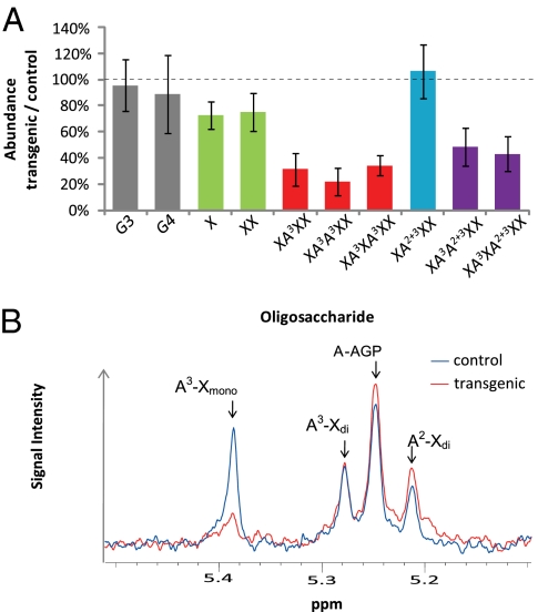
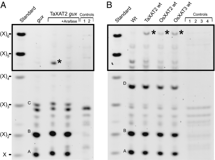
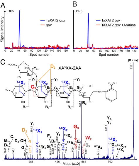
Xylan, a hemicellulosic component of the plant cell wall, is one of the most abundant polysaccharides in nature. In contrast to dicots, xylan in grasses is extensively modified by α-(1,2)- and α-(1,3)-linked arabinofuranose. Despite the importance of grass arabinoxylan in human and animal nutrition and for bioenergy, the enzymes adding the arabinosyl substitutions are unknown. Here we demonstrate that knocking-down glycosyltransferase (GT) 61 expression in wheat endosperm strongly decreases α-(1,3)-linked arabinosyl substitution of xylan. Moreover, heterologous expression of wheat and rice GT61s in Arabidopsis leads to arabinosylation of the xylan, and therefore provides gain-of-function evidence for α-(1,3)-arabinosyltransferase activity. Thus, GT61 proteins play a key role in arabinoxylan biosynthesis and therefore in the evolutionary divergence of grass cell walls.
Conflict of interest statement
Conflict of interest statement: The authors declare that a related patent application has been filed.
Figures
References
-
- Carpita NC. Structure and biogenesis of the cell walls of grasses. Annu Rev Plant Physiol Plant Mol Biol. 1996;47:445–476. - PubMed
-
- Scheller HV, Ulvskov P. Hemicelluloses. Annu Rev Plant Biol. 2010;61:263–289. - PubMed
-
- Ishii T. Isolation and characterization of a diferuloyl arabinoxylan hexasaccharide from bamboo shoot cell-walls. Carbohydr Res. 1991;219:15–22. - PubMed
-
- Sun R, Sun XF, Tomkinson J. Hemicelluloses: Science and Technology. Vol 864. Washington, DC: American Chemical Society; 2003. Hemicelluloses and their derivatives; pp. 2–22. ACS Symposium Series.
-
- Stone B, Morell MK. Carboyhdrates. In: Khan K, Shewry PR, editors. Wheat Chemistry and Technology. St Paul, Minnesota: AACC; 2009. pp. 299–362.
Publication types
MeSH terms
Substances
Grants and funding
- BB/C507561/1/BB_/Biotechnology and Biological Sciences Research Council/United Kingdom
- BB/F013434/1/BB_/Biotechnology and Biological Sciences Research Council/United Kingdom
- BB/G016240/1/BB_/Biotechnology and Biological Sciences Research Council/United Kingdom
- BB/F014295/1/BB_/Biotechnology and Biological Sciences Research Council/United Kingdom
LinkOut - more resources
Full Text Sources
Other Literature Sources
Molecular Biology Databases
Miscellaneous
