. 2012 Mar;32(5):992-1003.
doi: 10.1128/MCB.06665-11. Epub 2012 Jan 3.

A novel feedback loop regulates the response to endoplasmic reticulum stress via the cooperation of cytoplasmic splicing and mRNA translation

Affiliations

A novel feedback loop regulates the response to endoplasmic reticulum stress via the cooperation of cytoplasmic splicing and mRNA translation

Mithu Majumder et al. Mol Cell Biol. 2012 Mar.

Abstract

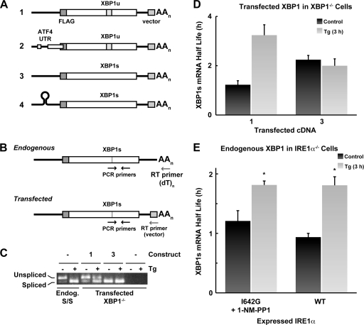
The accumulation of unfolded proteins in the endoplasmic reticulum (ER) triggers transcriptional and translational reprogramming. This unfolded protein response (UPR) protects cells during transient stress and can lead to apoptosis during prolonged stress. Two key mediators of the UPR are PKR-like ER kinase (PERK), which phosphorylates the α subunit of eukaryotic translation initiation factor 2 (eIF2α), resulting in decreased protein synthesis, and the α subunit of inositol-requiring enzyme 1 (IRE1α), which initiates cytoplasmic splicing of the mRNA encoding the transcription factor X-box binding protein 1 (XBP1). XBP1 induces transcription of genes involved in protein quality control. This report describes cross talk between these two pathways: phosphorylation of eIF2α was required for maximal induction of spliced XBP1 (XBP1s) protein levels via a mechanism that involved stabilization of XBP1s mRNA. By using mouse embryo fibroblasts deficient in UPR signaling pathways, we demonstrate that stress-induced stabilization of XBP1s mRNA requires cytoplasmic splicing of the mRNA and inhibition of its translation. Because the XBP1s protein promotes transcription of its own gene, the UPR-induced mRNA stabilization is part of a positive feedback loop that induces XBP1s protein accumulation and transcription of target genes during stress. We propose a model in which eIF2α phosphorylation-mediated control of mRNA turnover is a molecular switch that regulates the stress response transcription program and the ER's capacity for protein folding during stress.


Figures

Fig 1
Fig 1
XBP1s mRNA is stabilized after 3 h of the UPR in a process that requires translational repression. (A) S/S or A/A cells were incubated with Tg for the indicated times and analyzed for the indicated proteins by Western blotting. (B) S/S and A/A cells were incubated with Tg and/or Hip for the indicated times, and XBP1s mRNA levels were assessed by RT-qPCR. In the right-most sample, A/A cells were incubated for 6 h with Tg and Hip and then for 3 h with Tg only. (C) S/S and A/A cells were incubated with Tg for the indicated times, and the levels of XBP1u and XBP1s mRNAs were assessed by RT-qPCR. The graph shows the amount of XBP1s mRNA as a percentage of the total. (D) S/S and A/A cells were treated with Tg for 3 h and then treated with ActD and Tg for the indicated times, and XBP1s mRNA was quantified by RT-qPCR. Values were normalized to 18S rRNA and expressed as percentages of the levels before the addition of ActD.
Fig 2
Fig 2
Inhibition of protein synthesis in A/A cells stabilizes XBP1s mRNA. (A) A/A cells were incubated with Tg and Hip as indicated. After 3 h, ActD was added, and the XBP1s mRNA half-life was measured as described for Fig. 1. (B) S/S and A/A cells were treated as described for Fig. 1B, and XBP1s protein was quantified by Western blotting. Tubulin was measured as a loading control.
Fig 3
Fig 3
Splicing of XBP1 mRNA is required for increased stability during the UPR. (A) Expression plasmids used to analyze the role of mRNA splicing in the stabilization of XBP1s mRNA during the UPR. (B) Scheme for selective analysis of endogenous and exogenous XBP1s mRNAs by RT-PCR. To detect exogenous mRNAs, mRNAs were reverse transcribed using a primer complementary to vector sequences in the 3′-UTR. (C) XBP1−/− cells transfected with the indicated plasmids were cultured with Tg for 3 h. Splicing of endogenous mRNA from S/S cells and transgenic mRNAs from XBP1−/− cells were analyzed by RT-PCR and agarose gel electrophoresis. The specificity of the method for detecting exogenous mRNA was demonstrated by the lack of bands in untransfected cells. (D) XBP1−/− MEFs were transfected with the indicated constructs and treated with Tg for 3 h, and the half-life of XBP1s mRNA was measured as described for Fig. 1. (E) IRE1α−/− MEFs were transfected with either I642G or WT IRE1α expression plasmids. The former cells were cultured with 1-NM-PP1 overnight before the experiment. Cells were treated with Tg for 3 h, and the half-life of XBP1s mRNA was assessed as described for Fig. 1. *, response of Tg-treated cells was significantly different from the control value (P < 0.05).
Fig 4
Fig 4
mRNA poly(A) tail length does not affect the stabilization of XBP1s mRNA during the UPR. (A) Method for detecting mRNA poly(A) tail length. poly(G · I) tails were added to mRNAs followed by reverse transcription with a tail-specific primer. PCR was performed using a common upstream primer (3) and a downstream primer complementary to the added tail (1) or the 3′-UTR (2). The differences in the sizes of the products were due to the poly(A) tail. (B) S/S and A/A MEFs were cultured with Tg for the indicated times and analyzed as described for panel A. (C, D, and E) S/S cells were cultured with cordycepin for 2 h, followed by cordycepin and Tg for 3 h as indicated. (C) XBP1 mRNA splicing was assessed by RT-PCR; (D) poly(A) tail length was assessed; (E) XBP1s mRNA half-life was measured as described for Fig. 1.
Fig 5
Fig 5
Dephosphorylation of eIF2α during prolonged stress results in loss of XBP1s mRNA stabilization. (A) S/S and A/A cells were treated with Tg for the indicated times, and the incorporation of [35S]Met/Cys was measured. (B) S/S cells were cultured with Tg for 7 h, and the half-life of XBP1s mRNA was assessed as described for Fig. 1. *, result for Tg-treated cells was significantly different from the control value (P < 0.05). (C) GADD34−/− MEFs were cultured with Tg for the indicated times, and the XBP1s mRNA half-life was assessed as described for Fig. 1.
Fig 6
Fig 6
Decreased translational efficiency is required for stabilization of XBP1s mRNA during ER stress. (A) S/S cells were treated with Tg for the indicated times, and the distribution of XBP1s mRNA in polyribosomes was analyzed. (B) S/S cells were transfected with the indicated constructs from Fig. 3A and incubated with or without Tg for 3 h, and the splicing of XBP1 mRNA was assessed by RT-PCR and gel electrophoresis. (C) S/S cells were transfected with the indicated constructs (Fig. 3A), and the expression of Flag-XBP1s was detected by Western blotting using anti-FLAG antibody. (D) S/S cells were transfected and cultured as for panel B. (Left) The half-lives of the expressed XBP1s mRNAs were determined as described for Fig. 1. (Right) The distribution of XBP1s mRNA on polyribosome profiles was analyzed and used to calculate the percentage of the total signal in the polyribosome fraction. *, result for Tg-treated cells was significantly different from the control value (P < 0.05); **, result for control cells transfected with construct 2 was significantly different from cells with control cells with construct 1 (P < 0.05). A representative experiment for the distribution of XBP1s mRNA on polyribosome profiles is shown.
Fig 7
Fig 7
NMD-independent marking of the XBP1s mRNA during the UPR. (A to C) A/A cells were transfected with control siRNA or siRNA to UPF1. After 48 h, UPF1 protein was measured by Western blotting (A), XBP1s splicing was measured by RT-PCR (B), and the half-lives of XBP1s and DNABJ2 mRNAs were measured as described for Fig. 1. (D) S/S cells were treated with Tg and Hip as indicated, and the XBP1s mRNA half-life was evaluated after 8 h as described for Fig. 1.
Fig 8
Fig 8
Model for regulation of XBP1s mRNA stability during the UPR. In unstressed cells, basal IRE1α activity produces small amounts of XBP1s mRNA that are degraded rapidly. Induction of the UPR activates IRE1α, which induces XBP1 mRNA splicing and deposition of protein factors on the mRNA. Phosphorylation of eIF2α by PERK inhibits XBP1s mRNA translation. The combination of cytoplasmic splicing and translational inhibition leads to the reduced turnover of XBP1s mRNA. As the UPR progresses, GADD34 induces dephosphorylation of eIF2α and translational recovery. XBP1s protein is synthesized from the accumulated mRNA, which stimulates the transcription of XBP1 and its target genes. The loss of translational inhibition leads to rapid turnover of the XBP1s mRNA, limiting the accumulation of this protein.

References

    1. Adachi Y, et al. 2008. ATF6 is a transcription factor specializing in the regulation of quality control proteins in the endoplasmic reticulum. Cell Struct. Funct 33:75–89 - PubMed
    1. Anelli T, Sitia R. 2008. Protein quality control in the early secretory pathway. EMBO J. 27:315–327 - PMC - PubMed
    1. Barreau C, Paillard L, Osborne HB. 2005. AU-rich elements and associated factors: are there unifying principles? Nucleic Acids Res. 33:7138–7150 - PMC - PubMed
    1. Bertolotti A, Zhang Y, Hendershot LM, Harding HP, Ron D. 2000. Dynamic interaction of BiP and ER stress transducers in the unfolded- protein response. Nat. Cell Biol. 2:326–332 - PubMed
    1. Bevilacqua E, et al. 2010. eIF2α phosphorylation tips the balance to apoptosis during osmotic stress. J. Biol. Chem. 285:17098–17111 - PMC - PubMed

Publication types

MeSH terms

LinkOut - more resources