

High throughput, cell type-specific analysis of key proteins in human endometrial biopsies of women from fertile and infertile couples
- PMID: 22215622
- PMCID: PMC3279126
- DOI: 10.1093/humrep/der436
High throughput, cell type-specific analysis of key proteins in human endometrial biopsies of women from fertile and infertile couples
Abstract
Background: Although histological dating of endometrial biopsies provides little help for prediction or diagnosis of infertility, analysis of individual endometrial proteins, proteomic profiling and transcriptome analysis have suggested several biomarkers with altered expression arising from intrinsic abnormalities, inadequate stimulation by or in response to gonadal steroids or altered function due to systemic disorders. The objective of this study was to delineate the developmental dynamics of potentially important proteins in the secretory phase of the menstrual cycle, utilizing a collection of endometrial biopsies from women of fertile (n = 89) and infertile (n = 89) couples.
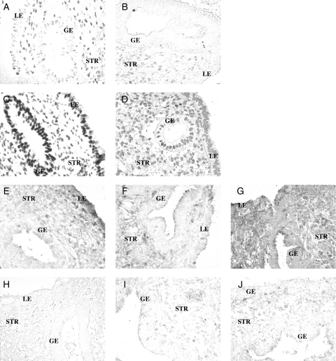
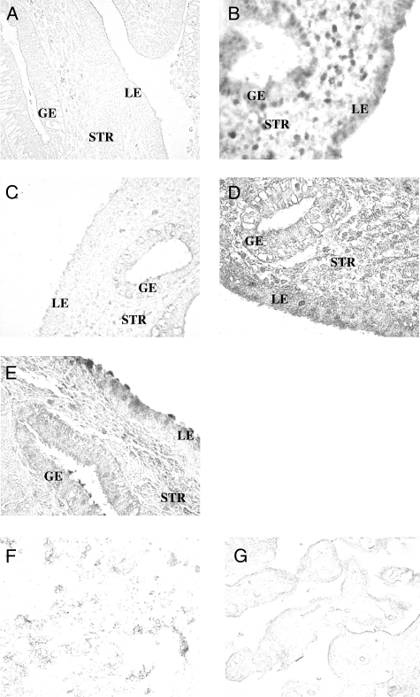
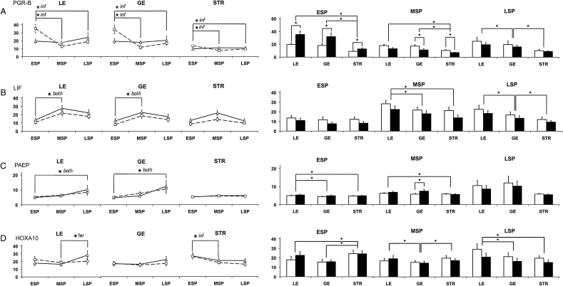
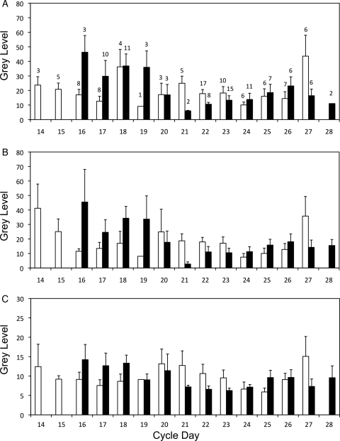
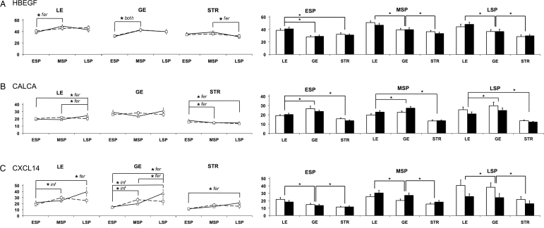
Methods and results: Progesterone receptor-B (PGR-B), leukemia inhibitory factor, glycodelin/progestagen-associated endometrial protein (PAEP), homeobox A10, heparin-binding EGF-like growth factor, calcitonin and chemokine ligand 14 (CXCL14) were measured using a high-throughput, quantitative immunohistochemical method. Significant cyclic and tissue-specific regulation was documented for each protein, as well as their dysregulation in women of infertile couples. Infertile patients demonstrated a delay early in the secretory phase in the decline of PGR-B (P < 0.05) and premature mid-secretory increases in PAEP (P < 0.05) and CXCL14 (P < 0.05), suggesting that the implantation interval could be closing early. Correlation analysis identified potential interactions among certain proteins that were disrupted by infertility.
Conclusions: This approach overcomes the limitations of a small sample number. Protein expression and localization provided important insights into the potential roles of these proteins in normal and pathological development of the endometrium that is not attainable from transcriptome analysis, establishing a basis for biomarker, diagnostic and targeted drug development for women with infertility.
Figures
Similar articles
-
Reduced homeobox protein MSX1 in human endometrial tissue is linked to infertility.Hum Reprod. 2016 Sep;31(9):2042-50. doi: 10.1093/humrep/dew143. Epub 2016 Jun 16. Hum Reprod. 2016. PMID: 27312535 Free PMC article.
-
Glycodelin in endometrial flushing fluid and endometrial biopsies from infertile and fertile women.Eur J Obstet Gynecol Reprod Biol. 2011 May;156(1):60-6. doi: 10.1016/j.ejogrb.2010.12.040. Epub 2011 Feb 2. Eur J Obstet Gynecol Reprod Biol. 2011. PMID: 21292383
-
HB-EGF but not amphiregulin or their receptors HER1 and HER4 is altered in endometrium of women with unexplained infertility.Reprod Sci. 2008 May;15(5):484-92. doi: 10.1177/1933719108314624. Reprod Sci. 2008. PMID: 18579857
-
Secretory proteins of the endometrium--potential markers for endometrial dysfunction.Baillieres Clin Obstet Gynaecol. 1989 Jun;3(2):271-91. doi: 10.1016/s0950-3552(89)80022-7. Baillieres Clin Obstet Gynaecol. 1989. PMID: 2482150 Review.
-
Endometrial proteins: a reappraisal.Hum Reprod. 1992 Jun;7 Suppl 1:31-8. doi: 10.1093/humrep/7.suppl_1.31. Hum Reprod. 1992. PMID: 1280281 Review.
Cited by
-
Endometrial shedding effect on conception and live birth in women with polycystic ovary syndrome.Obstet Gynecol. 2012 May;119(5):902-8. doi: 10.1097/AOG.0b013e31824da35c. Obstet Gynecol. 2012. PMID: 22525900 Free PMC article. Clinical Trial.
-
Changes in receptivity epithelial cell markers of endometrium after ovarian stimulation treatments: its role during implantation window.Reprod Health. 2015 May 17;12:45. doi: 10.1186/s12978-015-0034-7. Reprod Health. 2015. PMID: 25981399 Free PMC article.
-
Reduced expression of the epidermal growth factor signaling system in preeclampsia.Placenta. 2015 Mar;36(3):270-8. doi: 10.1016/j.placenta.2014.12.006. Epub 2014 Dec 27. Placenta. 2015. PMID: 25589361 Free PMC article.
-
Differential expression of serum glycodelin and insulin-like growth factor binding protein 1 in early pregnancy.Reprod Sci. 2013 Nov;20(11):1376-81. doi: 10.1177/1933719113485290. Epub 2013 Apr 12. Reprod Sci. 2013. PMID: 23585335 Free PMC article.
-
Cell signaling in trophoblast-uterine communication.Int J Dev Biol. 2014;58(2-4):261-71. doi: 10.1387/ijdb.140011da. Int J Dev Biol. 2014. PMID: 25023692 Free PMC article. Review.
References
-
- Aghajanova L, Velarde MC, Giudice LC. Altered gene expression profiling in endometrium: evidence for progesterone resistance. Semin Reprod Med. 2010;28:51–58. - PubMed
-
- Arimoto T, Katagiri T, Oda K, Tsunoda T, Yasugi T, Osuga Y, Yoshikawa H, Nishii O, Yano T, Taketani Y, et al. Genome-wide cDNA microarray analysis of gene-expression profiles involved in ovarian endometriosis. Int J Oncol. 2003;22:551–560. - PubMed
-
- Armant DR, Wang J, Liu Z. Intracellular signaling in the developing blastocyst as a consequence of the maternal-embryonic dialogue. Semin Reprod Med. 2000;18:273–287. - PubMed
-
- Arnett-Mansfield RL, DeFazio A, Mote PA, Clarke CL. Subnuclear distribution of progesterone receptors A and B in normal and malignant endometrium. J Clin Endocrinol Metab. 2004;89:1429–1442. - PubMed
Publication types
MeSH terms
Substances
Grants and funding
- R01 HD045966/HD/NICHD NIH HHS/United States
- HD38988/HD/NICHD NIH HHS/United States
- HD27011/HD/NICHD NIH HHS/United States
- HD38998/HD/NICHD NIH HHS/United States
- HD38992/HD/NICHD NIH HHS/United States
- ImNIH/Intramural NIH HHS/United States
- HD33172/HD/NICHD NIH HHS/United States
- HD38997/HD/NICHD NIH HHS/United States
- P50 HD055764/HD/NICHD NIH HHS/United States
- HD045966/HD/NICHD NIH HHS/United States
- U54HD40093/HD/NICHD NIH HHS/United States
- HD040093/HD/NICHD NIH HHS/United States
- HD39005/HD/NICHD NIH HHS/United States
- HD27049/HD/NICHD NIH HHS/United States
LinkOut - more resources
Full Text Sources
Medical
Research Materials
