. 2012 Feb 24;287(9):6941-8.
doi: 10.1074/jbc.M111.298703. Epub 2012 Jan 3.

Interleukin 33 as a mechanically responsive cytokine secreted by living cells

Affiliations

Interleukin 33 as a mechanically responsive cytokine secreted by living cells

Rahul Kakkar et al. J Biol Chem. .

Abstract

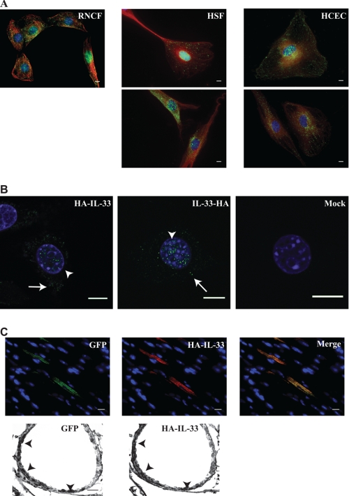
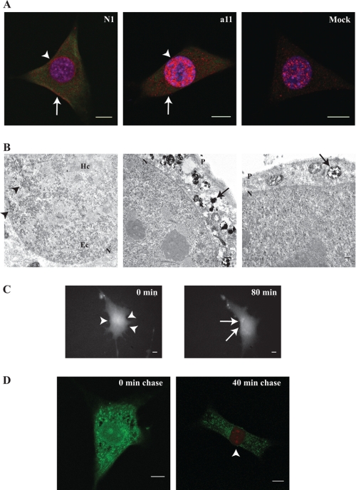
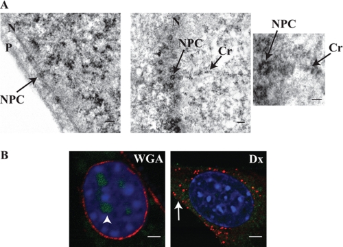
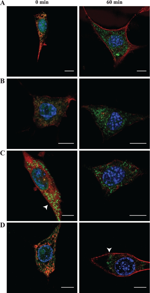
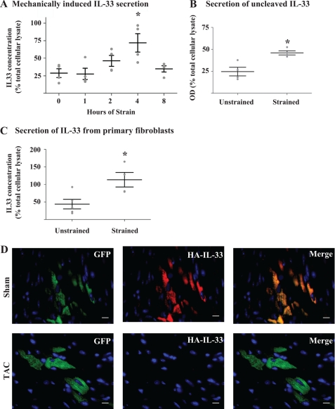
Interleukin 33 (IL-33), a member of the Interleukin 1 cytokine family, is implicated in numerous human inflammatory diseases such as asthma, atherosclerosis, and rheumatoid arthritis. Despite its pathophysiologic importance, fundamental questions regarding the basic biology of IL-33 remain. Nuclear localization and lack of an export signal sequence are consistent with the view of IL-33 as a nuclear factor with the ability to repress RNA transcription. However, signaling via the transmembrane receptor ST2 and documented caspase-dependent inactivation have suggested IL-33 is liberated during cellular necrosis to effect paracrine signaling. We determined the subcellular localization of IL-33 and tracked its intracellular mobility and extracellular release. In contrast to published data, IL-33 localized simultaneously to nuclear euchromatin and membrane-bound cytoplasmic vesicles. Fluorescent pulse-chase fate-tracking documented dynamic nucleo-cytoplasmic flux, which was dependent on nuclear pore complex function. In murine fibroblasts in vitro and in vivo, mechanical strain induced IL-33 secretion in the absence of cellular necrosis. These data document IL-33 dynamic inter-organelle trafficking and release during biomechanical overload. As such we recharacterize IL-33 as both an inflammatory as well as mechanically responsive cytokine secreted by living cells.


Figures

FIGURE 1.
FIGURE 1.
IL-33 simultaneously occupies multiple sub-cellular domains. A, IL-33 displays nuclear and cytoplasmic localization in wild type primary cells. The subcellular localization of IL-33 was interrogated by immunofluorescence epifluorescent microscopy utilizing two different anti-IL-33 antibodies. Rat neonatal cardiac fibroblasts (RNCF), human skin fibroblasts (HSF), and human coronary endothelial cells (HCEC) in culture were probed with either a commercially available anti-IL-33 antibody (upper panels) or our previously characterized anti-IL-33 antibody (lower panels). IL-33 signal is pseudocolored green, with anti-tubulin staining (denoting the cytoplasmic space) pseudocolored red. Note our anti-IL-33 antibody does not react with rat IL-33 under immunofluorescence protocols. Scale bars represent 10 μm. B, small epitope tags allow proper modeling of wild type IL-33 localization. In contrast to fluoroprotein-tagged IL-33, an HA tag at either the amino (HA-IL-33) or C (IL-33-HA) terminus permitted nuclear (arrowheads) and cytoplasmic (arrows) localization of stably expressed IL-33 in fibroblasts as seen by confocal microscopy in a pattern mimicking the wild type condition. Scale bars represent 10 μm. C, IL-33 occupies nuclear and cytoplasmic positions in vivo. Lentivirus harboring HA-IL-33 as well as a non-fusion GFP reporter was injected into the left ventricle (upper panels) or instilled into the trachea (lower panels) of anesthetized mice. Upon genomic integration and protein expression, cardiac myocytes revealed strong cytoplasmic HA-IL-33 staining (probed via an anti-HA antibody) in infected cells demarked by GFP expression. Respiratory epithelial cells of terminal bronchioles displayed nuclear and cytoplasmic immunohistochemical staining of HA-IL-33 in cells demarked by GFP expression (arrowheads). Images are representative of 2–3 animals per group and 6–10 microscopic fields per animal. Scale bars represent 10 μm.
FIGURE 2.
FIGURE 2.
IL-33 displays dynamic nucleo-cytoplasmic flux. A, tetracysteine-tagged IL-33 (TC-IL-33) displays nuclear and cytoplasmic localization. A fibroblast cell line stably expressing TC-IL-33 was probed with tetracysteine-avid fluorescein derivative and one of two anti-IL-33 antibodies (commercially available N1 or a11 raised by our laboratory). Confocal microscopy revealed nuclear (arrowheads) and cytoplasmic (arrows) localization of TC-IL-33 (pseudocolored green), similar to the pattern of wild-type IL-33 staining in mock-transfected cells (anti-IL33 antibody signal pseudocolored red). Note mock-transfected cells did not evidence fluorescein fluorescence. Scale bars represent 10 μm. B, cytoplasmic IL-33 is housed within vesicles. As the tetracysteine-avid resorufin derivative can render 3,3′-diaminobenzidine electron dense,(8) the subcellular localization of TC-IL-33 was interrogated by transmission electron microscopy. Nuclei displayed punctate electron densities (arrowheads) concentrated in areas of euchromatin (Ec). The cytoplasm evidenced electron densities within membrane-bound structures (arrows). P and N denote plasma and nuclear membranes, respectively. Hc indicates heterochromatin. Scale bars represent 1 μm. C, IL-33 displays dynamic nucleo-cytoplasmic flux. Fibroblasts stably expressing TC-IL-33 were pulsed with tetracysteine-avid fluorescein derivative and imaged by time-lapse epifluorescent microscopy. Shown are representative images at 0 and 80 min post-pulse. Over the chase period, a transposition of fluorescence from the nucleus (arrowheads) to the cytoplasmic space (arrows) is evident. A time-lapse movie may be viewed as part of the supplemental materials. Scale bars represent 10 μm. D, newly synthesized IL-33 molecules are nuclear in their localization. Pulse-chase-pulse experiments were performed by exposing fibroblasts stably expressing TC-IL-33 to a primary application of tetracysteine-avid fluorescein derivative followed by secondary resorufin derivative exposure after a variable chase period. By this method, all existent IL-33 molecules at the time of the initial pulse were rendered fluorescein-positive. Molecules synthesized after the primary pulse were non-fluorescent until application of the resorufin derivative at the end of the chase period, allowing differential fluorescence of molecules synthesized at different times. Cells treated with resorufin derivative without a chase period did not evidence red fluorescence in any subcellular compartment. Cells permitted a 40-min chase period evidenced fluorescein fluorescence in the cytoplasm and resorufin fluorescence in the nucleus (arrowhead). Scale bars represent 10 μm.
FIGURE 3.
FIGURE 3.
Nucleo-cytoplasmic shift of IL-33 is dependent on the nuclear pore complex. A, IL-33 localizes to nuclear pore complexes. High-magnification transmission electron microscopy of fibroblasts stably expressing TC-IL-33 exposed to the tetracysteine-avid resorufin-derivative displayed electron densities highlighting nuclear pore complexes (NPC). The left panel shows the complex in cross section. The middle panel depicts the complex en face whereby IL-33 molecules can be seen within the pore channel itself (enlarged in right panel). In these images, IL-33 can also be seen localizing to a chromatin strand (Cr). P and N denote plasma and nuclear membranes respectively. Scale bars represent 1 μm. B, normal nuclear pore complex function is obligatory for nuclear efflux of IL-33. Fibroblasts stably expressing TC-IL-33 were microinjected with either Texas-red conjugated wheat germ agglutinin (WGA) to block nuclear pore molecular transit or Texas-red conjugated dextran (Dx) as control. Cells were then treated with the tetracysteine-avid fluorescein derivative and allowed a 60-min chase period. In the left panel, Texas-red wheat germ agglutinin can be seen binding the plasma and nuclear membranes (pseudocolored red). Clear retention of nuclear fluorescein-labeled IL-33 can be seen (pseudocolored green, arrowhead). In contrast, control cells injected with Texas-red dextran (right panel) evidence efflux of IL-33 from the nucleus into the cytoplasm (arrow). Scale bars represent 5 μm.
FIGURE 4.
FIGURE 4.
IL-33 translocation depends on an intact microtubule network, and is ATP dependent. A–C, kinetics of IL-33 nucleo-cytoplasmic flux is enhanced upon microtubule disruption. A fibroblast cell line stably expressing TC-IL-33 was pulsed with tetracysteine-avid fluorescein derivative and allowed a chase period of 0 or 60 min (left and right panels, respectively) under variable conditions of chemical cytoskeletal disruption. A denotes control condition. B depicts experiments conducted in the presence of latrunculin B, to disrupt the actin cytoskeleton. C depicts experiments conducted in the presence of nocodazole, to disrupt the microtubule network. Under conditions of microtubule disruption, cytoplasmic vesicular IL-33 can be seen in the absence of a chase period (arrowhead). D, cytoplasmic transit of vesicular IL-33 is ATP dependent. Tetracysteine-avid fluorescein derivative pulse-chase was conducted under conditions of cellular ATP depletion (glucose free DMEM supplemented with 2-deoxy-d-glucose and 6 mm sodium azide). At the end of a 60-min chase period, punctate fluorescein staining can be seen relegated to a perinuclear position (arrowhead) rather than distributed throughout the cytoplasm as is seen in the control condition (panel A). Fluorescein staining is depicted in green. Phalloidin-based actin (latrunculin and ATP-depletion experiments) or anti-tubulin (nocodazole experiments) staining is depicted in red. Scale bars represent 10 μm.
FIGURE 5.
FIGURE 5.
Biomechanical strain induces IL-33 secretion from living cells. A, biomechanical strain induces IL-33 secretion. Fibroblasts stably expressing TC-IL-33 were subjected to 1 Hz cyclic 8% biaxial stretch for the number of hours indicated. Extracellular media was assayed via ELISA, and raw optical density values converted to IL-33 concentration via a standard curve followed by normalization to IL-33 in total cellular lysate. At 4 h, a 3.9-fold increase in optical density was measured (p = 0.012 by ANOVA; *, p < 0.01 for 0 versus 4 h by Bonferroni post-test; n = 4 per time point; whiskers represent mean and S.E.). B, IL-33 is released in full-length form. TC-IL-33 expressing cells were subjected to cyclic biaxial stretch for 4 h or left unstrained as control. Culture media was sampled, and TC-IL-33 was captured in a microplate coated with an anti-C-terminal IL-33 antibody. N-terminal tetracysteine fluorescence was induced by exposure to resorufin derivative dye. Raw optical density was normalized to the signal from total cellular lysate. A 1.8-fold increase in fluorescence was noted under conditions of strain compared with control (*, p = 0.008; n = 4 per condition; whiskers represent mean and S.E.). C, IL-33 is released from primary cells upon application of mechanical strain. Primary human skin fibroblasts were subjected to cyclic biaxial stretch for 4 h or left unstrained as control. Extracellular media was assayed via ELISA, and raw optical density values converted to IL-33 concentration via a standard curve followed by normalization to IL-33 in total cellular lysate. A 3.0-fold increase in optical density was noted in the context of strain (*, p = 0.02, n = 4–5 per condition; whiskers represent mean and S.E.). D, IL-33 is lost from cells subjected to biomechanical overload in vivo. Lentiviral particles housing HA-IL-33 and a non-fusion GFP reporter were injected into the left ventricle of anesthetized mice. Seven days later, mice underwent sham surgery (upper panels) or 2-h transaortic constriction (TAC, lower panels) to induce acute pressure overload of the left ventricle. Cardiac sections of sham hearts revealed strong HA-IL-33 staining (probed via an anti-HA antibody) in infected cells demarked by GFP expression. In hearts subjected to TAC, HA-IL-33 immunofluorescence was absent from GFP positive cells. Images are representative of 2–3 animals per group and 6–10 microscopic fields per animal. Scale bars represent 10 μm.

References

    1. Haraldsen G., Balogh J., Pollheimer J., Sponheim J., Küchler A. M. (2009) Interleukin-33 - cytokine of dual function or novel alarmin? Trends Immunol. 30, 227–233 - PubMed
    1. Carriere V., Roussel L., Ortega N., Lacorre D. A., Americh L., Aguilar L., Bouche G., Girard J. P. (2007) IL-33, the IL-1-like cytokine ligand for ST2 receptor, is a chromatin-associated nuclear factor in vivo. Proc. Natl. Acad. Sci. U.S.A. 104, 282–287 - PMC - PubMed
    1. Lingel A., Weiss T. M., Niebuhr M., Pan B., Appleton B. A., Wiesmann C., Bazan J. F., Fairbrother W. J. (2009) Structure of IL-33 and its interaction with the ST2 and IL-1RAcP receptors–insight into heterotrimeric IL-1 signaling complexes. Structure 17, 1398–1410 - PMC - PubMed
    1. Schmitz J., Owyang A., Oldham E., Song Y., Murphy E., McClanahan T. K., Zurawski G., Moshrefi M., Qin J., Li X., Gorman D. M., Bazan J. F., Kastelein R. A. (2005) IL-33, an interleukin-1-like cytokine that signals via the IL-1 receptor-related protein ST2 and induces T helper type 2-associated cytokines. Immunity 23, 479–490 - PubMed
    1. Cayrol C., Girard J. P. (2009) The IL-1-like cytokine IL-33 is inactivated after maturation by caspase-1. Proc. Natl. Acad. Sci. U.S.A. 106, 9021–9026 - PMC - PubMed

Publication types

MeSH terms