. 2012 Feb 17;287(8):5301-9.
doi: 10.1074/jbc.M111.291666. Epub 2012 Jan 3.

AMP is an adenosine A1 receptor agonist

Affiliations

AMP is an adenosine A1 receptor agonist

Joseph E Rittiner et al. J Biol Chem. .

Abstract

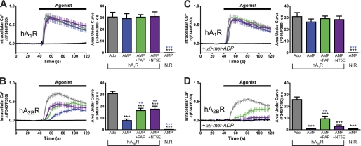
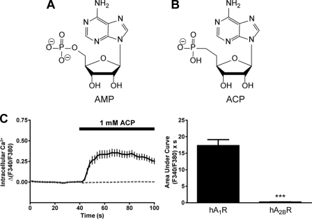
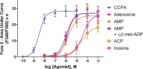
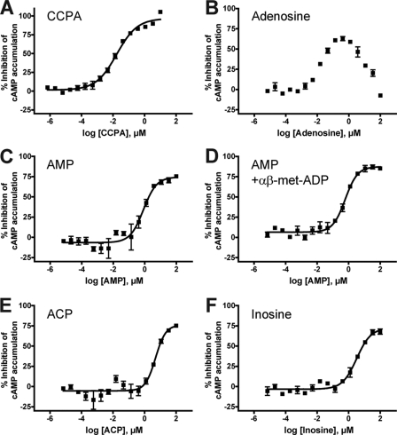
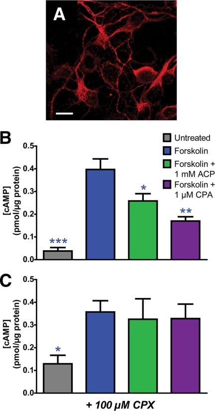
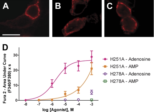
Numerous receptors for ATP, ADP, and adenosine exist; however, it is currently unknown whether a receptor for the related nucleotide adenosine 5'-monophosphate (AMP) exists. Using a novel cell-based assay to visualize adenosine receptor activation in real time, we found that AMP and a non-hydrolyzable AMP analog (deoxyadenosine 5'-monophosphonate, ACP) directly activated the adenosine A(1) receptor (A(1)R). In contrast, AMP only activated the adenosine A(2B) receptor (A(2B)R) after hydrolysis to adenosine by ecto-5'-nucleotidase (NT5E, CD73) or prostatic acid phosphatase (PAP, ACPP). Adenosine and AMP were equipotent human A(1)R agonists in our real-time assay and in a cAMP accumulation assay. ACP also depressed cAMP levels in mouse cortical neurons through activation of endogenous A(1)R. Non-selective purinergic receptor antagonists (pyridoxalphosphate-6-azophenyl-2',4'-disulfonic acid and suramin) did not block adenosine- or AMP-evoked activation. Moreover, mutation of His-251 in the human A(1)R ligand binding pocket reduced AMP potency without affecting adenosine potency. In contrast, mutation of a different binding pocket residue (His-278) eliminated responses to AMP and to adenosine. Taken together, our study indicates that the physiologically relevant nucleotide AMP is a full agonist of A(1)R. In addition, our study suggests that some of the physiological effects of AMP may be direct, and not indirect through ectonucleotidases that hydrolyze this nucleotide to adenosine.


Figures

FIGURE 1.
FIGURE 1.
AMP directly activates hA1R, whereas AMP activates hA2BR indirectly via ectonucleotidase-catalyzed hydrolysis to adenosine. A–D, calcium mobilization responses in HEK293 cells expressing (A and C) Gqi ± hA1R or (B and D) Gqs ± hA2BR. Ado, adenosine; N. R., no receptor. C and D, cells were incubated with the competitive NT5E inhibitor αβ-met-ADP (10 μm) for 3 min and then stimulated with 1 mm agonist in the presence of 10 μm αβ-met-ADP. Black, 1 mm AMP in the absence of a transfected adenosine receptor, but in the presence of the respective chimeric G protein. AUC measurements extended for 1 min from agonist addition. Paired t tests were used to compare AUC data. Black asterisks, statistically significant difference when compared with adenosine stimulation. Blue asterisks, statistically significant difference when compared with AMP stimulation (in receptor-expressing cells). *, p < 0.05, **, p < 0.005, ***, p < 0.0005. All data are the average of two experiments performed in duplicate. n = 20–74 cells per condition. All data, including calcium traces, are presented as means ± S.E.
FIGURE 2.
FIGURE 2.
Non-hydrolyzable AMP analog activates hA1R but not hA2BR. A and B, structures of AMP (A) and the non-hydrolyzable analog ACP (B) at physiological pH. C, calcium mobilization responses elicited by 1 mm ACP in HEK293 cells expressing (solid line) hA1R + Gqi or (dashed line) hA2BR + Gqs. Cells expressing Gqi alone did not respond to ACP. AUC measurements extended for 1 min from agonist addition. Paired t tests were used to compare AUC data, ***, p < 0.0005. Data are the average of one (hA2BR) or two (hA1R) experiments performed in duplicate. n = 28–73 cells per condition. All data, including calcium traces, are presented as means ± S.E.
FIGURE 3.
FIGURE 3.
Dose-response curves for adenosine, AMP, and related analogs at hA1R. Calcium mobilization was measured in HEK293 cells expressing hA1R + Gqi and stimulated with the indicated compounds. For the AMP + αβ-met-ADP condition, cells were incubated with 10 μm αβ-met-ADP for 3 min and then stimulated with AMP in the presence of 10 μm αβ-met-ADP. AUC measurements extended for 1 min from agonist addition. All experiments were performed in duplicate. n = 19–45 cells per condition. All data are presented as means ± S.E.
FIGURE 4.
FIGURE 4.
Adenosine, AMP, and related analogs inhibit cAMP accumulation in hA1R-expressing cells. A–F, HEK293T cells were co-transfected with a vector encoding hA1R and GloSensor 22F plasmid and then stimulated with CCPA (A), adenosine (B), AMP (C) or AMP in the presence of 10 μm αβ-met-ADP (D), ACP (E), or inosine (F). Cells were incubated with test compound for 10 min, and then 175 nm (−)-isoproterenol was added for 7 min to stimulate cAMP accumulation. Following incubation, GloSensor cAMP reagent was added, and luminescence was measured. Data were normalized such that 100% inhibition is equal to the response at the maximal concentration of CCPA and 0% is equal to the response from isoproterenol alone. All experiments were performed in duplicate. All data are presented as means ± S.D.
FIGURE 5.
FIGURE 5.
ACP inhibits cAMP accumulation in mouse embryonic cortical neurons. Embryonic cortical neurons were dissociated and plated at about embryonic day 16.5. A, confocal image of cortical neurons immunostained with an anti-A1R antibody. Scale bar = 10 μm. B and C, after 1 day in vitro, neurons were incubated for 30 min with 1 mm ACP or 1 μm cyclopentyladenosine (CPA) in the absence (B) or presence (C) of 100 μm CPX. Neurons were then stimulated with 10 μm forskolin for 15 min, washed, and lysed. Cell lysates were then applied to a cAMP ELISA assay according to the manufacturer's instructions. cAMP concentrations were normalized to total protein using a BCA protein assay. Asterisks, statistically significant difference when compared with forskolin stimulation alone. *, p < 0.05, **, p < 0.005, ***, p < 0.0005. Data are the average of four (− CPX) or two (+ CPX) experiments performed in duplicate. All data are presented as means ± S.E.
FIGURE 6.
FIGURE 6.
Expression and activity of hA1R point mutants. A–C, confocal images of HEK293 cells expressing wild-type hA1R (A), hA1R-H251A (B), or hA1R-H278A (C) and immunostained with anti-A1R antibodies. Untransfected HEK293 cells in the same field of view were not immunostained, confirming antibody specificity. Scale bar = 10 μm. D, calcium mobilization in HEK293 cells co-expressing the indicated hA1R mutant and Gqi and then stimulated with increasing concentrations of the indicated compounds. For all conditions, cells were incubated with 10 μm αβ-met-ADP for 3 min and then stimulated with agonist in the presence of 10 μm αβ-met-ADP. AUC measurements extended for 1 min from agonist addition. All experiments were performed in duplicate, except H278A-AMP, which was performed in triplicate. n = 17–49 cells per condition. All data are presented as means ± S.E.

References

    1. Burnstock G. (2007) Physiology and pathophysiology of purinergic neurotransmission. Physiol. Rev. 87, 659–797 - PubMed
    1. Jacobson K. A., Gao Z. G. (2006) Adenosine receptors as therapeutic targets. Nat. Rev. Drug Discov. 5, 247–264 - PMC - PubMed
    1. Inbe H., Watanabe S., Miyawaki M., Tanabe E., Encinas J. A. (2004) Identification and characterization of a cell surface receptor, P2Y15, for AMP and adenosine. J. Biol. Chem. 279, 19790–19799 - PubMed
    1. Abbracchio M. P., Burnstock G., Boeynaems J. M., Barnard E. A., Boyer J. L., Kennedy C., Miras-Portugal M. T., King B. F., Gachet C., Jacobson K. A., Weisman G. A. (2005) The recently deorphanized GPR80 (GPR99) proposed to be the P2Y15 receptor is not a genuine P2Y receptor. Trends Pharmacol. Sci. 26, 8–9 - PMC - PubMed
    1. Qi A. D., Harden T. K., Nicholas R. A. (2004) GPR80/99, proposed to be the P2Y15 receptor activated by adenosine and AMP, is not a P2Y receptor. Purinergic Signal 1, 67–74 - PMC - PubMed

Publication types

MeSH terms