. 2012 Mar 23;287(13):10639-10649.
doi: 10.1074/jbc.M111.300681. Epub 2012 Jan 3.

Ringo/cyclin-dependent kinase and mitogen-activated protein kinase signaling pathways regulate the activity of the cell fate determinant Musashi to promote cell cycle re-entry in Xenopus oocytes

Affiliations

Ringo/cyclin-dependent kinase and mitogen-activated protein kinase signaling pathways regulate the activity of the cell fate determinant Musashi to promote cell cycle re-entry in Xenopus oocytes

Karthik Arumugam et al. J Biol Chem. .

Abstract

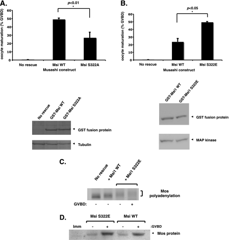
Cell cycle re-entry during vertebrate oocyte maturation is mediated through translational activation of select target mRNAs, culminating in the activation of mitogen-activated protein kinase and cyclin B/cyclin-dependent kinase (CDK) signaling. The temporal order of targeted mRNA translation is crucial for cell cycle progression and is determined by the timing of activation of distinct mRNA-binding proteins. We have previously shown in oocytes from Xenopus laevis that the mRNA-binding protein Musashi targets translational activation of early class mRNAs including the mRNA encoding the Mos proto-oncogene. However, the molecular mechanism by which Musashi function is activated is unknown. We report here that activation of Musashi1 is mediated by Ringo/CDK signaling, revealing a novel role for early Ringo/CDK function. Interestingly, Musashi1 activation is subsequently sustained through mitogen-activated protein kinase signaling, the downstream effector of Mos mRNA translation, thus establishing a positive feedback loop to amplify Musashi function. The identified regulatory sites are present in mammalian Musashi proteins, and our data suggest that phosphorylation may represent an evolutionarily conserved mechanism to control Musashi-dependent target mRNA translation.


Figures

FIGURE 1.
FIGURE 1.
Musashi1 undergoes stimulus-dependent phosphorylation. A, Musashi is required to mediate oocyte maturation. Musashi function was attenuated by injection of antisense DNA oligonucleotides targeting the endogenous Musashi1 and Musashi2 mRNAs as previously described (13). Oocytes were reinjected with water (no rescue) or RNA encoding GST-tagged wild-type Musashi1 and either left untreated (−prog) or stimulated with progesterone (+prog). The ability of the ectopic Musashi to rescue cell cycle progression was assessed (% GVBD). Results from two independent experiments are shown. A GST Western blot confirmed expression of the ectopic Musashi1 protein (arrowhead, lower panel). B, conservation of Ser-322 and flanking amino acids in vertebrate Musashi proteins is shown. The amino acid sequence flanking the identified serine phosphorylation site (bold) is conserved in human (hu), mouse (mu), Xenopus (Xl), and zebrafish (dr) Musashi1 and Musashi2 isoforms but not in C. elegans (Ce) or Drosophila (Dm). C, Musashi is phosphorylated on Ser-322 in response to progesterone stimulation. Musashi (WT) or a non-phosphorylatable mutant Musashi (S332A) were expressed in oocytes as GST fusion proteins and then stimulated with progesterone. Protein and RNA samples were prepared at the indicated time points. At 5.5 h, 50% of the injected oocytes had completed GVBD, and they were segregated based on whether they had (+) or had not (−) completed GVBD. A Western blot of oocyte lysates with antibody specific to the Ser-322 phosphorylated form of Musashi1 demonstrates progesterone-stimulated phosphorylation that is not observed in the S322A mutant Musashi (upper panel). A GST Western blot shows equivalent levels of the expressed proteins (lower panel). D, the Musashi mRNA target, Mos, is activated coincident with Musashi1 Ser-322 phosphorylation. Total RNA from samples shown in C was analyzed for polyadenylation of the endogenous Mos mRNA. Increased PCR product size indicates polyadenylation, and this initiates ∼3 h after progesterone stimulation (asterisk). E, endogenous Musashi1 was phosphorylated on Ser-322 in response to progesterone stimulation. Uninjected oocytes were stimulated with progesterone, and Western blots of protein lysate prepared at the indicated time points were assayed with antisera specific for Ser-322 phosphorylated Musashi1 as described in C. Quantitation of maximum progesterone-induced changes in phospho-Musashi1 levels normalized to tubulin from the same samples revealed a 1.25 ± 0.11-fold increase (Student's t test; p < 0.05, n = 4) relative to levels in immature oocytes.
FIGURE 2.
FIGURE 2.
Musashi1 Ser-322 phosphorylation facilitates oocyte maturation and target mRNA translational activation. A, inhibition of Musashi1 Ser-322 phosphorylation attenuates oocyte maturation. Oocytes were injected with antisense oligonucleotides to ablate endogenous Musashi function and subsequently reinjected with water (no rescue), GST-tagged wild-type Musashi1 (Msi WT), or the non-phosphorylatable mutant Musashi (Msi S322A) and scored for progesterone-dependent maturation when 50% of Msi WT expressing oocytes reached GVBD. Error bars represent S.E. from four independent experiments (p < 0.01, Student's t test). The Msi WT and S322A proteins were expressed to equivalent levels in the rescue assay as assessed by GST Western blotting (lower panel). A Western blot of the same lysates with tubulin antiserum confirmed equivalent protein loading. B, mutational mimicry of Musashi1 Ser-322 phosphorylation accelerates oocyte maturation. Oocytes were injected with Musashi antisense oligonucleotides as described in A and subsequently reinjected with water (No rescue), GST wild-type Musashi1 (Msi WT), or the phosphomimetic mutant Musashi (Msi S322E), and progesterone-dependent maturation was scored when 50% of Musashi S322E-expressing oocytes reached GVBD. Error bars represent S.E. from three independent experiments (p < 0.05, Student's t test). The Msi WT and S322E proteins were expressed to equivalent levels in the rescue assay as assessed by GST Western blot (lower panel). Western blot of the same lysates with MAP kinase antiserum confirmed equivalent protein loading. C, mutational mimicry of Musashi1 Ser-322 phosphorylation accelerates translational activation of the Mos mRNA. Total RNA was isolated from oocytes expressing GST Msi WT or the GST Msi S332E mutant protein described in B. Samples were prepared when 50% of Msi S322E oocytes reached GVBD and segregated based on whether they had or had not completed GVBD. Samples from time-matched Msi WT and water-injected (No rescue) were also prepared, and endogenous Mos mRNA polyadenylation was assessed. D, mutational mimicry of Musashi Ser-322 phosphorylation enhances Mos protein accumulation. Protein lysates were isolated from oocytes expressing GST Msi WT or the GST Msi S322E protein, and Mos protein levels were determined by Western blot. Samples were prepared when 50% of the injected oocytes reached GVBD and segregated based on whether they had or had not completed GVBD. Note the Msi S322E-expressing oocytes display higher Mos protein levels despite being harvested 30 min before Msi WT oocytes. Imm, immature oocytes.
FIGURE 3.
FIGURE 3.
Musashi function is necessary for Ringo-induced early class mRNA translational activation. A, inhibition of Musashi blocks Ringo-induced translational activation of the Mos mRNA. Immature oocytes were injected with RNA encoding the dominant inhibitory Musashi (N-Msi) or with water, incubated to allow expression of the N-Msi protein, and subsequently reinjected with RNA encoding Ringo. Total RNA was prepared at various times after Ringo RNA injection, and progression through maturation (GVBD) was assessed. The time required for 50% of the oocyte population to reach GVBD was significantly delayed in N-Msi-expressing oocytes (7 versus 4 h). Polyadenylation of the endogenous Mos mRNA was assessed as described in the legend to Fig. 2C. In the N-Msi expressing oocytes, Ringo did not induce polyadenylation of the Mos mRNA, and deadenylation of the Mos mRNA was observed. B, Ringo/CDK induces Musashi Ser-322 phosphorylation. Immature oocytes were injected with RNA encoding GST tagged Musashi1 and incubated overnight. The oocytes were then left untreated (Imm) or re-injected with RNA encoding Ringo or with inactive Ringo D83A, which does not activate CDK activity, as indicated, and time-matched protein lysates were prepared when 50% of Ringo-injected oocytes completed GVBD. Ringo-injected oocytes were segregated based on whether they had (+) or had not completed GVBD (−). Ringo D83A-injected oocytes did not mature. Western blotting was performed with appropriate antisera to analyze phosphorylation of Musashi1 Ser-322, phosphorylation (activation) of MAP kinase, ectopic Ringo protein expression, and expression of GST-Musashi1 protein as indicated. C, Ringo is required for progesterone-stimulated Musashi Ser-322 phosphorylation. Immature oocytes were co-injected with RNA encoding GST-Musashi1 and either control antisense oligonucleotides (Con AS) or antisense oligonucleotides targeting the endogenous Ringo mRNA (Ringo AS). The injected oocytes were then left untreated (Imm) or stimulated with progesterone (+prog). Oocytes were collected when the 50% of the control population had reached GVBD (3.5 h) and were segregated along with time-matched Ringo AS injected oocytes, and protein lysates were analyzed for Musashi1 Ser-322 phosphorylation and GST-Musashi expression by Western blot. No maturation of Ringo AS injected oocytes was observed at the time points analyzed.
FIGURE 4.
FIGURE 4.
Ringo/CDK and MAP kinase signaling pathways direct Musashi phosphorylation on Ser-322. A, MAP kinase signaling induces Musashi1 Ser-322 phosphorylation independently of CDK. Immature oocytes were injected with RNA encoding GST-tagged Musashi1 in the presence or absence of RNA encoding the CDK inhibitor Wee107 and incubated overnight. Oocytes were then split into pools and left untreated, injected with RNA encoding vRaf, or stimulated with progesterone as indicated. Progesterone- and vRaf-stimulated oocytes were segregated when the populations reached 50% GVBD (− and + GVBD, respectively). Protein lysates were analyzed by Western blot for Musashi1 phosphorylation on Ser-322, MAP kinase phosphorylation (indicative of activation), Cdc2 phosphorylation (indicative of inactive cyclin B/CDK), and relative GST-Musashi1 expression. B, progesterone-stimulated Musashi1 Ser-322 phosphorylation is mediated by both MAP kinase-dependent and MAP kinase-independent signaling. Immature oocytes were injected with RNA encoding GST-Musashi1 and incubated overnight, then treated with UO126 to inhibit MAP kinase signaling (+) or DMSO vehicle control (−) for 30 min before progesterone addition. Time-matched protein lysates were prepared when 50% of the control oocytes reached GVBD. Control oocytes were segregated based on whether they had (+) or had not (−) completed GVBD. Lysates were analyzed by Western blot for phosphorylation of Musashi1 on Ser-322, phosphorylation (activation) of MAP kinase, total MAP kinase, and expression of GST-Musashi1 in the same samples. C, shown is a schematic illustrating relevant progesterone-dependent signaling events impinging upon early class Musashi-mediated, mRNA translation before MPF (cyclin B/CDK) activation and oocyte GVBD. The points of experimental manipulation are shown along with the deduced Musashi amplification loops. For the sake of clarity, the complex roles of CPEB are not indicated in the diagram (“Discussion”).
FIGURE 5.
FIGURE 5.
Phosphorylation of Ser-322 and Ser-297 mediates Musashi activation. A, shown is conservation of Ser-297 and flanking amino acids in vertebrate Musashi proteins. Schematic alignment of Ser-297 (bold) and flanking amino acids in a range of organisms (see the legend to Fig. 1B) is shown. B, inhibition of Musashi1 Ser-297 and Ser-322 phosphorylation attenuates oocyte maturation. Oocytes were injected with antisense oligonucleotides to ablate endogenous Musashi function and subsequently reinjected with water (No rescue), GST-tagged wild-type Musashi1 (Msi WT), or the non-phosphorylatable double mutant Musashi (Msi S297A/S322A) and scored for progesterone-dependent maturation when 50% of Msi WT expressing oocytes reached GVBD. Error bars represent S.E. from four independent experiments (p < 0.01, Student's t test). The Msi WT and Msi S297A/S322A proteins were expressed to equivalent levels in the rescue assay as assessed by GST Western blotting (lower panel). C, inhibition of Musashi1 Ser-297 and Ser-322 phosphorylation attenuates Mos mRNA polyadenylation. Oocytes were treated with Musashi antisense oligonucleotides and subsequently reinjected as described in B. Total RNA samples were prepared when 50% of Msi WT oocytes reached GVBD and segregated based on whether they had not or had completed GVBD (− and +, respectively). Samples from time-matched water injected (No rescue) as well as Msi S297A/S322A-injected oocytes were also prepared, and endogenous Mos mRNA polyadenylation was assessed. Uninjected oocyte samples were also analyzed as a positive control for progesterone-induced Mos mRNA polyadenylation. An increase in size of the PCR product in progesterone-treated oocytes is indicative of polyadenylation. Imm, immature oocyte. D, mutational mimicry of Musashi1 Ser-297 and Ser-322 phosphorylation accelerates oocyte maturation. Oocytes were injected with Musashi antisense oligonucleotides as described in A and subsequently reinjected with water (No rescue), GST wild-type Musashi1 (Msi WT), or the phosphomimetic double mutant Musashi (Msi S297E/S322E), and progesterone-dependent maturation was scored when 50% of Musashi S297E/S322E-expressing oocytes reached GVBD. Error bars represent S.E. from three independent experiments (p < 0.05, Student's t test). The Msi WT and Msi S297E/S322E proteins were expressed to equivalent levels in the rescue assay as assessed by GST Western blot (lower panel).
FIGURE 6.
FIGURE 6.
Phospho-mimetic mammalian Musashi1 displays abrogated target mRNA repression in NIH3T3 cells. A, expressed murine Musashi1 is phosphorylated on Ser-337 in progesterone-stimulated oocytes. Immature Xenopus oocytes were injected with RNA encoding GST-tagged murine Musashi1 (mMsi1) and were stimulated with progesterone (prog) or left untreated (Imm). When 50% of the progesterone-stimulated oocyte population completed GVBD, oocytes were segregated (− or + GVBD) and analyzed by Western blot with phospho-Ser-322 Musashi-specific antiserum and with GST antiserum to show levels of the expressed protein (GST mMsi1). The progesterone-dependent appearance of Ser-337-phosphorylated mammalian Musashi1 is coincident with a gel mobility shift of the expressed Musashi protein. B, phospho-mimetic Musashi1 S337E has reduced ability to repress translation of target mRNAs in NIH3T3 cells. MBE-regulated firefly luciferase reporter mRNA was co-transfected with plasmids encoding GST (Control), wild-type murine Musashi1, or murine Musashi1 S337E, and after 48 h cell incubation luciferase activity was assessed and normalized to co-expressed Renilla luciferase (20). Luciferase activity in cells expressing wild-type mMusashi1 and mMusashi1 S337E was compared with luciferase activity in cell expressing the GST tag alone (set at 100%). Error bars represent S.E. from three independent experiments (p < 0.001, Student's t test). C, GST and tubulin Western blot of lysates in B show equivalent protein expression. D, the levels of Firefly luciferase reporter mRNA under the control of a Musashi-binding element containing 3′ UTR (Fluc-MBE) were determined using semiquantitative PCR. Total RNA was prepared from the indicated co-transfection conditons, and Fluc-MBE reporter mRNA was PCR-amplified for different cycle numbers as indicated. The PCR products were visualized after separation through a 2% agarose gel. No significant differences in stability of the Fluc-MBE construct (arrowhead) were detected in cells expressing the GST moiety alone (Control), GST wild-type Musashi1, or GST Musashi1 S337E. m, indicates DNA marker lane.

References

    1. Deng J., Carbajal L., Evaul K., Rasar M., Jamnongjit M., Hammes S. R. (2009) Nongenomic steroid-triggered oocyte maturation of mice and frogs. Steroids 74, 595–601 - PMC - PubMed
    1. Downs S. M. (2010) Regulation of the G2/M transition in rodent oocytes. Mol. Reprod. Dev. 77, 566–585 - PMC - PubMed
    1. Bettegowda A., Smith G. W. (2007) Mechanisms of maternal mRNA regulation. Implications for mammalian early embryonic development. Front. Biosci. 12, 3713–3726 - PubMed
    1. Radford H. E., Meijer H. A., de Moor C. H. (2008) Translational control by cytoplasmic polyadenylation in Xenopus oocytes. Biochim. Biophys. Acta 1779, 217–229 - PMC - PubMed
    1. Charlesworth A., Wilczynska A., Thampi P., Cox L. L., MacNicol A. M. (2006) Musashi regulates the temporal order of mRNA translation during Xenopus oocyte maturation. EMBO J. 25, 2792–2801 - PMC - PubMed

Publication types

MeSH terms

LinkOut - more resources