Comparative Study
. 2012 Feb;59(2):256-64.
doi: 10.1161/HYPERTENSIONAHA.111.182170. Epub 2012 Jan 3.

Transcriptionally active syncytial aggregates in the maternal circulation may contribute to circulating soluble fms-like tyrosine kinase 1 in preeclampsia

Affiliations
Comparative Study

Transcriptionally active syncytial aggregates in the maternal circulation may contribute to circulating soluble fms-like tyrosine kinase 1 in preeclampsia

Augustine Rajakumar et al. Hypertension. 2012 Feb.

Abstract

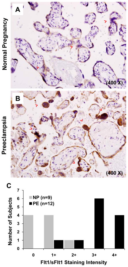
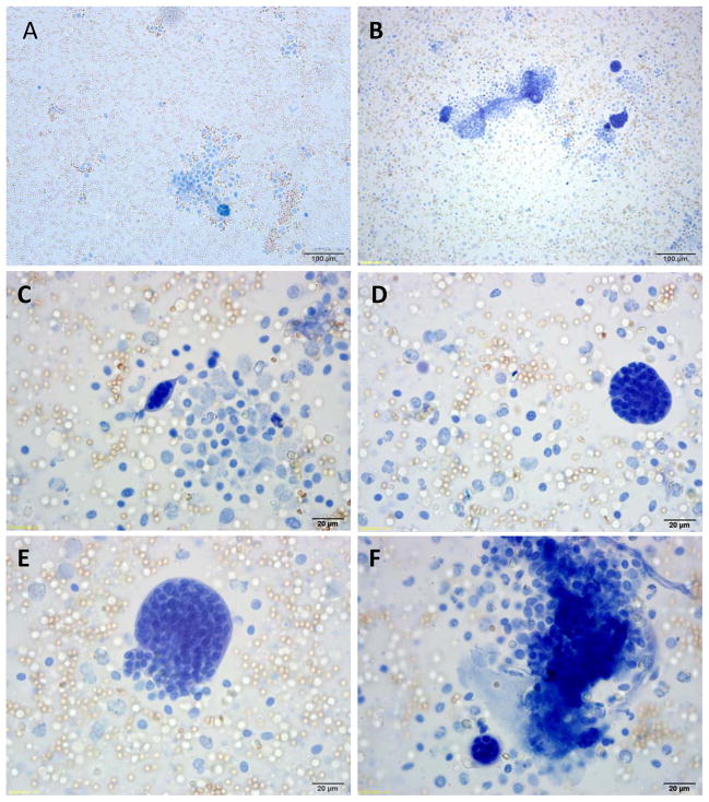
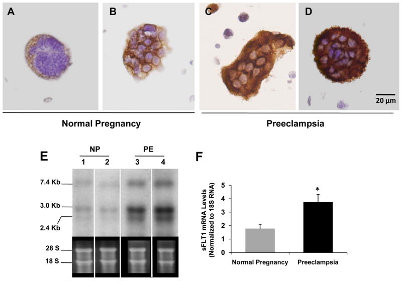
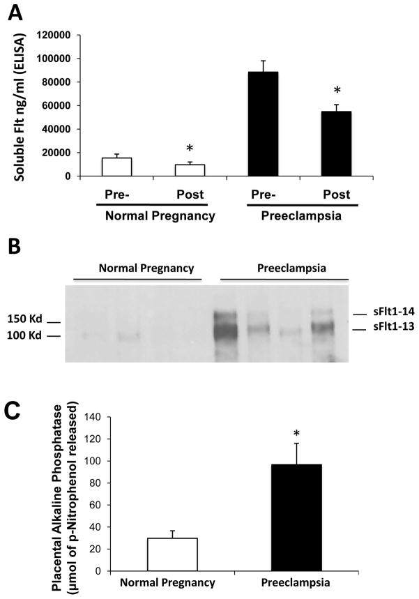
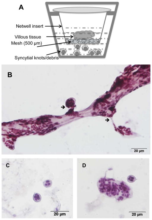
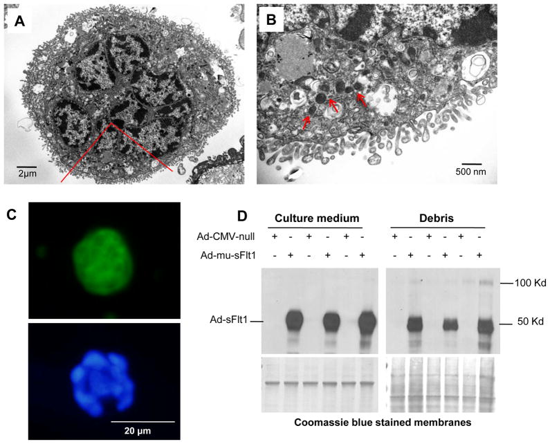
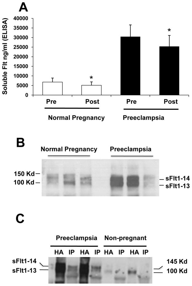
The cardinal manifestations of the pregnancy-specific disorder preeclampsia, new-onset hypertension, and proteinuria that resolve with placental delivery have been linked to an extracellular protein made by the placenta, soluble fms-like tyrosine kinase 1 (sFlt1), that injures the maternal vasculature. However, the mechanisms by which sFlt1, which is heavily matrix bound, gain access to the systemic circulation remain unclear. Here we report that the preeclamptic placenta's outermost layer, the syncytiotrophoblast, forms abundant "knots" that are enriched with sFlt1 protein. These syncytial knots easily detach from the syncytiotrophoblast, resulting in free, multinucleated aggregates (50-150 μm diameter) that are loaded with sFlt1 protein and mRNA, are metabolically active, and are capable of de novo gene transcription and translation. At least 25% of the measurable sFlt1 in the third-trimester maternal plasma is bound to circulating placental microparticles. We conclude that detachment of syncytial knots from the placenta results in free, transcriptionally active syncytial aggregates that represent an autonomous source of sFlt1 delivery into the maternal circulation. The process of syncytial knot formation, shedding of syncytial aggregates, and appearance of placental microparticles in the maternal circulation appears to be greatly accelerated in preeclampsia and may contribute to the maternal vascular injury that characterizes this disorder.


Figures

Figure 1
Figure 1. Immunohistochemistry for sFlt1 expression in normal and preeclamptic placentae
Immunohistochemical staining and analysis of placental tissues from normal pregnancy (n=9) and preeclampsia (n=12) for Flt/sFlt1 expression were performed. Panel A and B show a representative staining of normal and preeclamptic placenta, at term, respectively. The red arrowheads represent syncytial knots. Magnification 400X. Panel C shows a graphical representation of the quantitation of the Flt/sFlt1 staining.
Figure 2
Figure 2. Analyses of placental washes from normal and preeclamptic placentae
Representative photomicrograph of Trypan blue staining of the contents of the placental washes obtained from a normal (panel A) and preeclamptic women (panel B). Panels C – F show different sized syncytial aggregates in preeclamptic placental effluents. Red blood cells and leukocytes can be seen in the background.
Figure 3
Figure 3. Expression of sFlt1 mRNA and protein in syncytial debris obtained from placental washes
Panels A to D show sFlt1 staining by IHC of the syncytial knots obtained from placental washes from two normal pregnant women (A and B) and from two preeclamptic women (C and D). Scale bar 20 μm in all the panels. RNA obtained from placental washes of normal pregnant (NP) (n=5) and preeclamptic women (PE) (n=6) was analyzed by Northern blot. A representative blot from two samples of each category is shown in panel E and the quantitation in a graph in panel F. *p=<0.05 by ANOVA.
Figure 4
Figure 4. Microparticle associated sFlt1 in the culture medium of placental villous explants
Placental villous explants were cultured as described in materials and methods and supernatant was analyzed for sFlt1 expression. High-speed centrifugation reduced sFlt1 by ~30% in placental explant conditioned medium. The pre- and post-spin sFlt1 levels measured by ELISA are shown in Panel A. sFlt1 in 100K pellets was detected by Western blot analyses (Panel B). The quantitation of placental alkaline phosphatase (PlAP) by densitometry from the Western blots is shown in panel C. *p<0.05 by ANOVA.
Figure 5
Figure 5. Characterization of placental syncytial aggregates using ex vivo organ cultures
Panel A is a schematic of the ex vivo placental organ cultures on Netwell inserts. Panel B shows the generation of syncytial knots of different sizes from the main villous tissue. Arrow shows the break point of multinucleated aggregates from the villous tissue. Panel C–D shows representative high power images of individual multi-nucleated aggregates that have separated from the villous tissue.
Figure 6
Figure 6. Syncytial aggregates exhibit transcriptional and translational capacities
(A–B). Electron micrograph of multinucleated aggregate shows of microvillous cell membrane and abundant cytoplasmic organelles (Panel A). A region marked in red is magnified to show mitochondria (Panel B) with red arrowheads. Panel C. GFP expression in a syncytial aggregate infected with GFP adenovirus is shown in the upper panel. Nuclear co-localization was done with DAPI blue, bottom panel. Panel D. Syncytial aggregates were infected with adenovirus carrying truncated sFlt1 protein and the conditioned medium was analyzed by Western blot for expression of sFlt1 protein.
Figure 7
Figure 7. Microparticle associated sFlt1 in plasma of pregnant women
Panel A. Plasma samples from normal pregnant (n=12) and preeclamptic women (n=15) at term show an approximate 25% reduction in circulating sFlt1 levels after ultracentrifugation. Panel B is a representative western blot analyses for sFlt1 protein expression in the 100K pellets of the plasma obtained from normal and preeclamptic women. Panel C demonstrates that sFlt1 containing microparticles also express syncytin. Plasma samples from preeclamptic patients (n=2) and non-pregnant women (n=2) were subjected to ultracentrifugation. The 100K pellet was precipitated using heparin-agarose (HA) or using syncytin antibody (IP) and Western blot performed with antibody directed against the N-terminus of Flt1. * p< 0.05 by ANOVA.

Comment in

References

    1. Redman CW, Sargent IL. Latest advances in understanding preeclampsia. Science. 2005;308:1592–1594. - PubMed
    1. Sibai B, Dekker G, Kupferminc M. Pre-eclampsia. Lancet. 2005;365:785–799. - PubMed
    1. Ahmad S, Ahmed A. Elevated placental soluble vascular endothelial growth factor receptor-1 inhibits angiogenesis in preeclampsia. Circ Res. 2004;95:884–891. - PubMed
    1. Chaiworapongsa T, Romero R, Espinoza J, Bujold E, Mee Kim Y, Goncalves LF, Gomez R, Edwin S. Evidence supporting a role for blockade of the vascular endothelial growth factor system in the pathophysiology of preeclampsia. Am J Obstet Gynecol. 2004;190:1541–1547. - PubMed
    1. Levine RJ, Maynard SE, Qian C, Lim KH, England LJ, Yu KF, Schisterman EF, Thadhani R, Sachs BP, Epstein FH, Sibai BM, Sukhatme VP, Karumanchi SA. Circulating angiogenic factors and the risk of preeclampsia. N Engl J Med. 2004;350:672–683. - PubMed

Publication types

Substances