

MRI of the cervical spine with neck extension: is it useful?
- PMID: 22215879
- PMCID: PMC3587072
- DOI: 10.1259/bjr/94315429
MRI of the cervical spine with neck extension: is it useful?
Abstract
Objectives: Standard MRI of the cervical spine is performed in a different anatomical position to that utilised for traditional contrast myelography. Those well practised in myelography are familiar with the considerable changes in configuration of the bony and soft tissues of the cervical spine that may occur with changes in the degree of neck flexion and extension. We set out to compare the findings in a select group of patients with myeloradiculopathy who had undergone myelography and MRI in both standard and neck-extended positions. These findings were correlated with the clinical status.
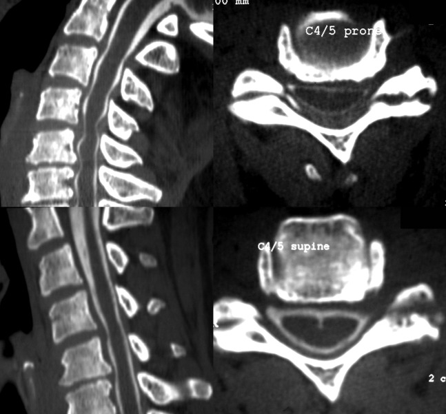
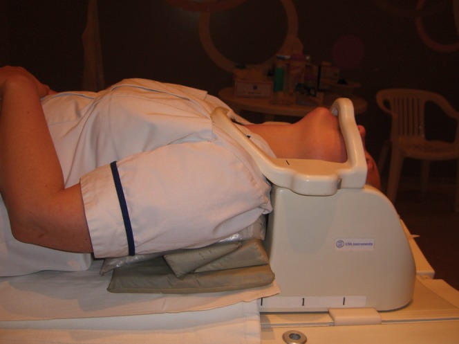
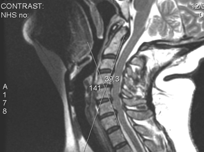
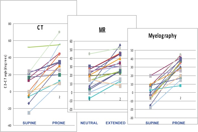
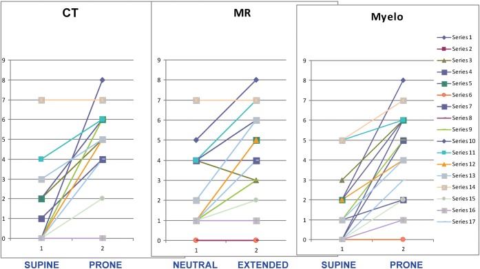
Methods: 29 patients underwent myelography with CT (CTM) and MRI in neutral and neck-extended positions. The imaging was assessed for the degree of cord compression and neural foraminal narrowing, quantified using a simple grading scheme suitable for routine clinical practice. The degree of neck extension was assessed using an angular measurement.
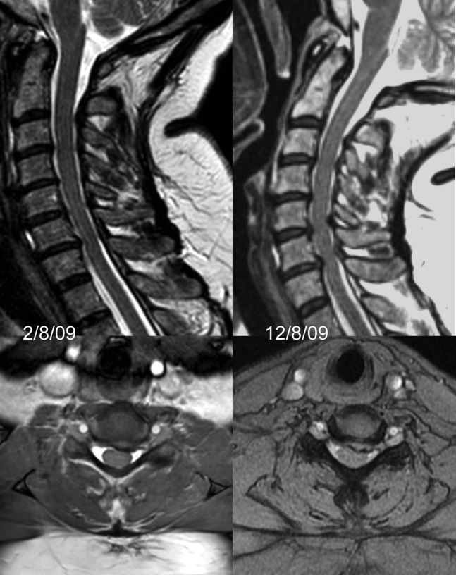
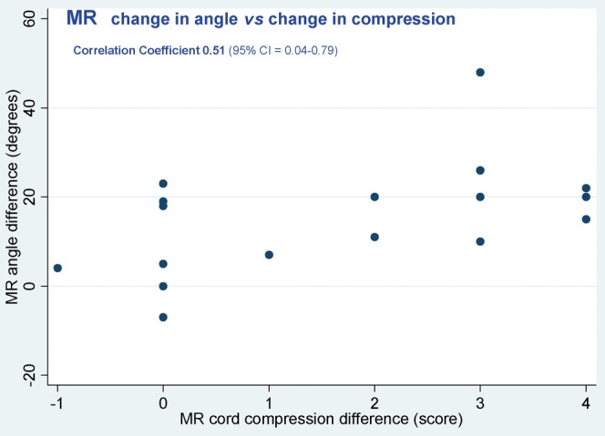
Results: For both CTM and MRI, scanning with the neck extended significantly increases the severity of cord compression compared with the standard supine position, to a degree similar to that shown during conventional prone myelography. The degree of perceived cord compression is related to the degree of neck extension achieved. Correlation of standard MRI findings and the clinical level of radiculopathy is poor. This correlation improves when the neck is extended.
Conclusions: The most appropriate position for routine MRI of the cervical spine in degenerative disease remains unknown, but in selected patients imaging with the neck extended may provide important additional information.
Figures
Similar articles
-
Clinical usefulness of CT-myelogram comparing with the MRI in degenerative cervical spinal disorders: is CTM still useful for primary diagnostic tool?J Spinal Disord Tech. 2009 Jul;22(5):353-7. doi: 10.1097/BSD.0b013e31817df78e. J Spinal Disord Tech. 2009. PMID: 19525791
-
Comparison of computed tomography myelography and magnetic resonance imaging in the evaluation of cervical spondylotic myelopathy and radiculopathy.Spine (Phila Pa 1976). 1999 Sep 1;24(17):1781-5. doi: 10.1097/00007632-199909010-00006. Spine (Phila Pa 1976). 1999. PMID: 10488507
-
Comparison of CT myelography performed in the prone and supine positions in the detection of cervical spinal stenosis.Clin Radiol. 2001 Jan;56(1):35-9. doi: 10.1053/crad.2000.0569. Clin Radiol. 2001. PMID: 11162695 Clinical Trial.
-
[Imaging cervical myelo- and radiculopathy].Radiologe. 2006 Nov;46(11):993-1000. doi: 10.1007/s00117-005-1251-9. Radiologe. 2006. PMID: 16133405 Review. German.
-
Diagnosis of nerve root compression. Myelography, computed tomography, and MRI.Orthop Clin North Am. 1992 Jul;23(3):405-19. Orthop Clin North Am. 1992. PMID: 1620535 Review.
Cited by
-
Kinetic DTI of the cervical spine: diffusivity changes in healthy subjects.Neuroradiology. 2016 Sep;58(9):929-35. doi: 10.1007/s00234-016-1709-7. Epub 2016 Jun 8. Neuroradiology. 2016. PMID: 27278377
-
Evaluation of Dynamic Foraminal Stenosis with Positional MRI in Patients with C6 Radiculopathy-Mimicking Pain: A Prospective Radiologic Cohort Study.Biomed Res Int. 2022 Jun 9;2022:1385387. doi: 10.1155/2022/1385387. eCollection 2022. Biomed Res Int. 2022. PMID: 35722464 Free PMC article.
-
Comparison of two MR grading systems for correlation between grade of cervical neural foraminal stenosis and clinical manifestations.Br J Radiol. 2016 Jun;89(1062):20150971. doi: 10.1259/bjr.20150971. Epub 2016 Mar 23. Br J Radiol. 2016. PMID: 27007709 Free PMC article.
-
Can microstructural MRI detect subclinical tissue injury in subjects with asymptomatic cervical spinal cord compression? A prospective cohort study.BMJ Open. 2018 Apr 13;8(4):e019809. doi: 10.1136/bmjopen-2017-019809. BMJ Open. 2018. PMID: 29654015 Free PMC article.
-
Prone extension views in cervical MRI: A case-driven Novel approach.Heliyon. 2023 Dec 8;10(1):e23251. doi: 10.1016/j.heliyon.2023.e23251. eCollection 2024 Jan 15. Heliyon. 2023. PMID: 38163148 Free PMC article.
References
-
- Graham CB, Wippold FJ, Bae KT, Pilgram TK, Shaibani A, Kido DK. Comparison of CT myelography performed in the prone and supine positions in the detection of cervical spinal stenosis. Clin Rad 2001;56:35–9 - PubMed
-
- Benini A. Clinical features of cervical root compression C5–C8 and their variations. Nearal Orthop 1987;4:74–88
-
- Modic MT, Masaryk TJ, Mulopulos GP, Bundschuh C, Han JS, Bohlman H. Cervical radiculopathy: prospective evaluation with surface coil MR imaging, CT with metrizamide and metrizamide myelography. Radiology 1986;161:753–9 - PubMed
-
- Takahashi M, Yamashita Y, Sakamoto Y, Kojima R. Chronic cervical cord compression: clinical significance of increased signal intensity on MR images. Radiology 1989;173:219–24 - PubMed
Publication types
MeSH terms
LinkOut - more resources
Full Text Sources
Medical
