

Investigating the relationship between virtual cystoscopy image quality and CT slice thickness
- PMID: 22215882
- PMCID: PMC3587099
- DOI: 10.1259/bjr/99567374
Investigating the relationship between virtual cystoscopy image quality and CT slice thickness
Abstract
Objective: To investigate the effect of reconstruction slice thickness on image quality at CT virtual cystoscopy (VC).
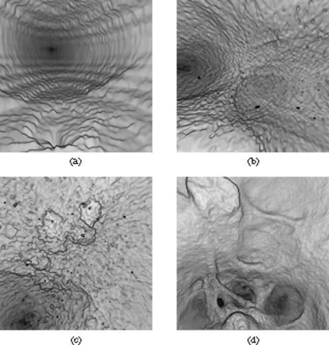
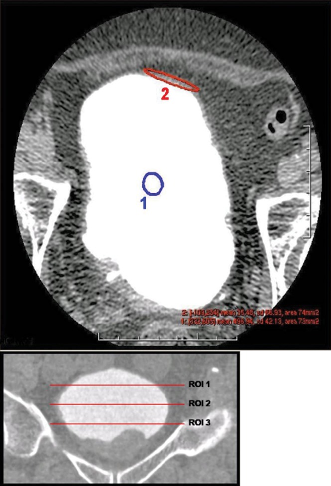
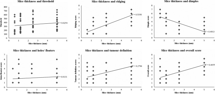
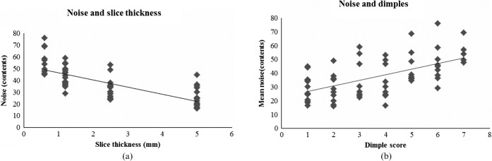
Methods: Pelvic CT examinations in bladder cancer patients were reconstructed at different slice thicknesses (0.6-5 mm) and intervals, and resulting VC images assessed. Quality indicators were ridging, holes, floaters and dimpling artefacts, tumour definition, and an overall score, ranked 1 (best) to 7 (worst). CT number and standard deviation (SD) for bladder contents and bladder wall were recorded. The mean SD was used as a measure of noise, and the contrast-to-noise ratio (CNR) was calculated as the CT number difference between them divided by the average image noise. The mean CNR across the three levels was used for analysis. Each qualitative image quality measure was compared with CT number, noise and CNR measurements.
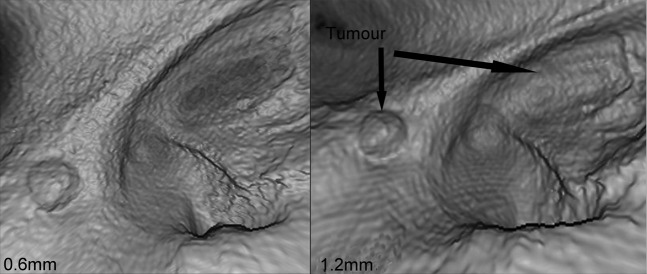
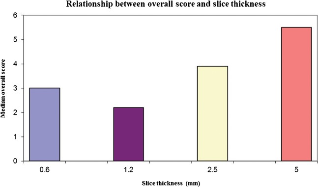
Results: Dimpling artefacts increased with thinner slice reconstruction and correlated with increased noise, often resulting in poor tumour definition. The best overall image quality score was seen for VC images reconstructed at 1.2 mm slice thickness, probably because of the competing effects of spatial resolution and CNR.
Conclusion: A slice thickness reconstruction <1.2 mm does not provide for better image quality at VC owing to the presence of increased noise.
Figures
References
-
- Collado A, Chechile GE, Salvador J, Vicente J. Early complications of endoscopic treatment for superficial bladder tumors. J Urol 2000;164:1529–32 - PubMed
-
- Hollenbeck BK, Miller DC, Taub D, Dunn RL, Khuri SF, Henderson WG, et al. Risk factors for adverse outcomes after transurethral resection of bladder tumors. Cancer 2006;106:1527–35 - PubMed
-
- Nieder AM, Meinbach DS, Kim SS, Soloway MS. Transurethral bladder tumor resection: intraoperative and postoperative complications in a residency setting. J Urol 2005;174:2307–9 - PubMed
-
- Levin B, Lieberman DA, McFarland B, Smith RA, Brooks D, Andrews KS, et al. Screening and surveillance for the early detection of colorectal cancer and adenomatous polyps, 2008: a joint guideline from the American Cancer Society, the US Multi-Society Task Force on Colorectal Cancer, and the American College of Radiology. CA Cancer J Clin 2008;58:130–60 - PubMed
-
- Kim JK, Park SY, Kim HS, Kim SH, Cho KS. Comparison of virtual cystoscopy, multiplanar reformation, and source CT images with contrast material-filled bladder for detecting lesions. AJR Am J Roentgenol 2005;185:689–96 - PubMed
