. 2011 Dec;7(12):e1002412.
doi: 10.1371/journal.pgen.1002412. Epub 2011 Dec 22.

A complex genomic rearrangement involving the endothelin 3 locus causes dermal hyperpigmentation in the chicken

Affiliations

A complex genomic rearrangement involving the endothelin 3 locus causes dermal hyperpigmentation in the chicken

Ben Dorshorst et al. PLoS Genet. 2011 Dec.

Abstract

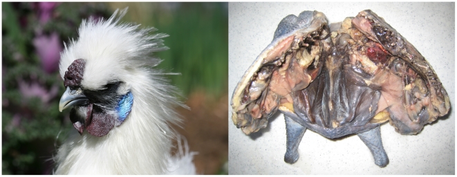
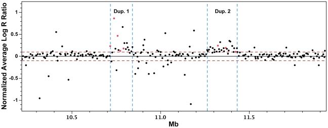
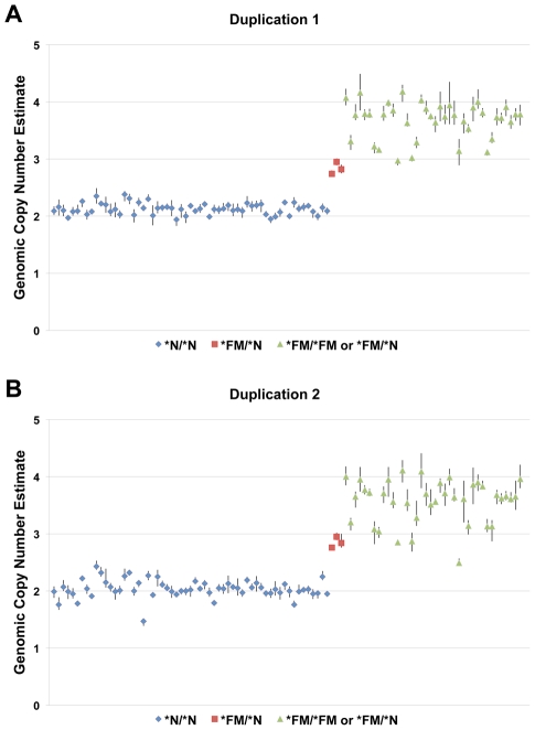
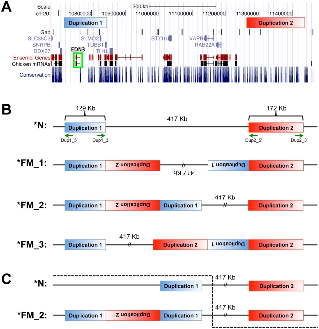
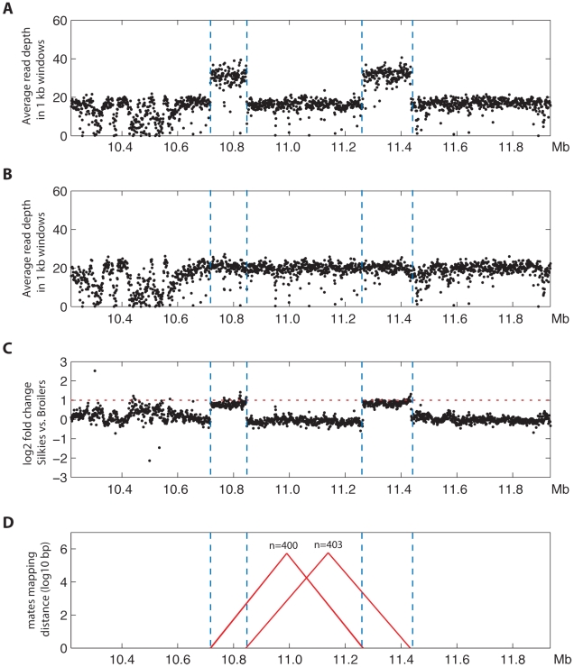
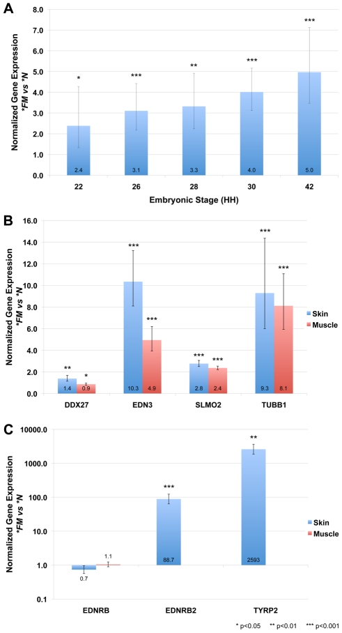
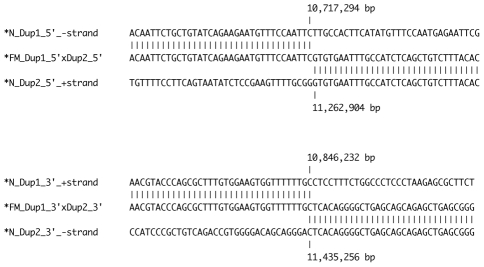
Dermal hyperpigmentation or Fibromelanosis (FM) is one of the few examples of skin pigmentation phenotypes in the chicken, where most other pigmentation variants influence feather color and patterning. The Silkie chicken is the most widespread and well-studied breed displaying this phenotype. The presence of the dominant FM allele results in extensive pigmentation of the dermal layer of skin and the majority of internal connective tissue. Here we identify the causal mutation of FM as an inverted duplication and junction of two genomic regions separated by more than 400 kb in wild-type individuals. One of these duplicated regions contains endothelin 3 (EDN3), a gene with a known role in promoting melanoblast proliferation. We show that EDN3 expression is increased in the developing Silkie embryo during the time in which melanoblasts are migrating, and elevated levels of expression are maintained in the adult skin tissue. We have examined four different chicken breeds from both Asia and Europe displaying dermal hyperpigmentation and conclude that the same structural variant underlies this phenotype in all chicken breeds. This complex genomic rearrangement causing a specific monogenic trait in the chicken illustrates how novel mutations with major phenotypic effects have been reused during breed formation in domestic animals.


Conflict of interest statement

The authors have declared that no competing interests exist.

Figures

Figure 1
Figure 1. The Silkie chicken displaying the Fibromelanosis phenotype.
An adult White Silkie Bantam chicken (left). Hyperpigmentation of the comb, wattle, face, and beak is clearly visible. A White Silkie Bantam (right) prepared in a typical manner for meat consumption, having being split down the spine with viscera removed. Intense pigmentation of internal connective tissue and the exterior skin is evident while muscle tissue remains normally pigmented.
Figure 2
Figure 2. Group-wise analysis of Log R ratio SNP data for the detection of copy number variations.
Log R ratio data from wild-type and FM individuals were partitioned based on FM genotype. The average of the wild-type group was subtracted from the average of the *FM group on an individual SNP basis and plotted by genomic base pair coordinate. Two distinct genomic regions with elevated Log R ratio values are evident (red dotted line = ±0.1). SNPs marked in red were always observed in the heterozygous state in FM individuals in both duplicated regions.
Figure 3
Figure 3. A two-fold increase in genomic copy number is associated with FM.
Genomic copy number was estimated using qPCR for a large panel of individuals with known skin pigmentation status. Panel A depicts a primer/probe set located within the first duplicated region and panel B depicts a primer/probe set located within the second duplication. Three known heterozygotes (red) show an estimated copy number of approximately 3 as compared to wild-type individuals (blue) with a copy number of 2. Individuals known to carry at least one *FM allele (green) cluster towards an estimated copy number of four, although it is evident that some heterozygotes are likely included in this group.
Figure 4
Figure 4. Genome view of duplicated regions and possible rearrangement scenarios.
(A) The location of the two duplicated regions is depicted in blue and red respectively. The location of EDN3 is outlined in green. Several other genes are located within the first duplicated region while no known coding elements are found within the second duplicated region. Image was generated with the UCSC Genome Browser (http://genome.ucsc.edu) using the May 2006 (WUGSC 2.1/galGal3) assembly. (B) The structural arrangement of the FM locus was tested with outward facing primers (green arrows) at the boundary of each duplicated region. The amplification pattern obtained using different primer combinations was consistent with three different rearrangement scenarios; *FM_1, *FM_2, and *FM_3 as compared to the wild-type *N arrangement. (C) A single individual in the backcross mapping population strongly supports the *FM_2 rearrangement scenario. This individual was a recombinant in the 417 kb single copy region as represented by the dashed line, possessing the alleles inherited from the *N founder prior to the crossover event and the alleles inherited from the *FM founder from the crossover onwards. This individual was phenotypically wild-type and had the normal copy number of both duplicated regions, all of which supports *FM_2 as the only possible arrangement of the *FM allele.
Figure 5
Figure 5. Massively parallel sequencing confirms the inverted duplication corresponding to the FM locus.
Average sequencing read depths in windows of 1 kb along the interval 10.218–11.935 Mb on chromosome 20 for (A) the Silkie pool and (B) the Broiler pool. (C) Log2 fold change values (normalized for read depth) between Silkie and Broiler pools showing that the interval within the two large duplications (area between blue vertical dotted lines) has approximately twice as high levels of sequence coverage in the Silkie pool than in the Broiler pool (2× higher levels indicated by the horizontal red dotted line). (D) The mate-pair information was used to plot all candidate structural variants in the region of interest. Candidate structural variants were defined as windows where at least 20% of the mate pairs had mapping distances exceeding six standard deviations above the average mapping distance for chromosome 20 and had mapping distances ranging ±1500 base pairs from the median distance observed for those exceeding 6 standard deviations. On the y-axis the size of candidate structural variants are presented in log10 base pairs and the x coordinates of connected colored lines indicate the genomic coordinates of the pairs supporting structural variants (red = mate-pairs map to different strands, which is indicative of an inversion). The number of mate-pairs supporting a feature is indicated above the feature.
Figure 6
Figure 6. Genomic rearrangement drives increased expression of genes located within the duplicated region in FM embryonic and adult tissues.
Gene expression analysis by SYBR Green qPCR of *FM (Silkie breed) tissue normalized to the *N (New Hampshire breed) and calibrated to glyceraldehyde 3-phosphate dehydrogenase (GAPDH). Error bars indicate 95% confidence intervals and significance thresholds are indicated. (A) Embryo tissue cross sections were collected at the level of the wing bud at the indicated stages. EDN3 is upregulated at all developmental stages assayed, with an increasing magnitude of differential expression in *FM tissue with age. (B) Genes located within the first duplicated region (EDN3, SLMO2 and TUBB1) are significantly increased in expression in adult skin and muscle tissue of *FM chickens. The expression of DDX27, located adjacent to but outside of the duplicated region, is also significantly differentially expressed, but does not show the same degree of upregulation as the genes within the duplication. (C) The expression of the EDN3 receptor EDNRB is not significantly different in *FM skin or muscle tissue when compared to *N tissue. However, the expression of the EDN3 receptor EDNRB2, expressed predominately by melanocytes, is highly upregulated in *FM skin tissue. A key component of the melanin biosynthesis pathway, TYRP2, is also highly upregulated in *FM skin tissue. EDNRB2 and TYRP2 expression was not detected in *N muscle tissue, so no comparison can be made to *FM skin tissue.
Figure 7
Figure 7. Sequence alignment of duplication junction points.
The junction of the 5′ portion of the duplicated regions was detected using primers Dup1_5' and Dup2_5' and the junction of the 3′ portion of each duplicated region was detected using primers Dup1_3' and Dup2_3'. No sequence homology, insertion or deletion was detected at the breakpoints except for the overlap of a single C nucleotide at the junction of the 3′ portion of the duplicated regions.

References

    1. Hutt FB. Genetics of the fowl. New York: McGraw-Hill; 1949. pp. 590.
    1. Faraco CD, Vaz SA, Pastor MV, Erickson CA. Hyperpigmentation in the Silkie fowl correlates with abnormal migration of fate-restricted melanoblasts and loss of environmental barrier molecules. Dev Dyn. 2001;220:212–225. - PubMed
    1. Kuklenski J. Uber das Vorkommen und die Verteilung des Pigmentes in den Organen und Geweben bei japanischen seiden Huhnern. Arch Micro Anat Entwickl. 1915;87:1–37.
    1. Smyth JR., Jr . Genetics of plumage, skin and eye pigmentation in chickens; In: Crawford RD, editor. Amsterdam; New York: Elsevier; 1990. pp. 109–167.
    1. Muroya S, Tanabe R, Nakajima I, Chikuni K. Molecular characteristics and site specific distribution of the pigment of the silky fowl. J Vet Med Sci. 2000;62:391–395. - PubMed

Publication types

MeSH terms