Review
. 2012:2012:589589.
doi: 10.1155/2012/589589. Epub 2011 Dec 14.

ER stress and apoptosis: a new mechanism for retinal cell death

Affiliations
Review

ER stress and apoptosis: a new mechanism for retinal cell death

Guangjun Jing et al. Exp Diabetes Res. 2012.

Abstract

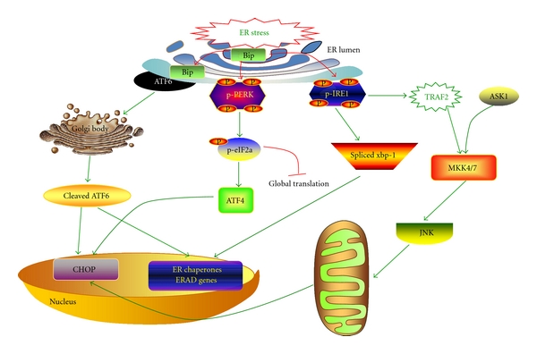
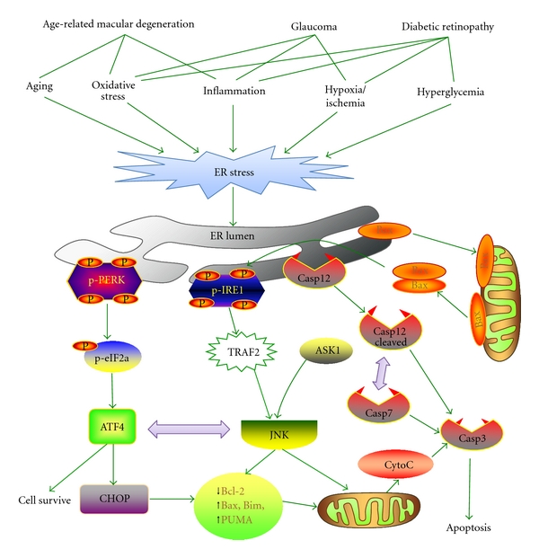
The endoplasmic reticulum (ER) is the primary subcellular organelle where proteins are synthesized and folded. When the homeostasis of the ER is disturbed, unfolded or misfolded proteins accumulate in the ER lumen, resulting in ER stress. In response to ER stress, cells activate a set of tightly controlled regulatory programs, known as the unfolded protein response (UPR), to restore the normal function of the ER. However, if ER stress is sustained and the adaptive UPR fails to eliminate unfolded/misfolded proteins, apoptosis will occur to remove the stressed cells. In recent years, a large body of studies has shown that ER stress-induced apoptosis is implicated in numerous human diseases, such as diabetes and neurogenerative diseases. Moreover, emerging evidence supports a role of ER stress in retinal apoptosis and cell death in blinding disorders such as age-related macular degeneration and diabetic retinopathy. In the present review, we summarize recent progress on ER stress and apoptosis in retinal diseases, focusing on various proapoptotic and antiapoptotic pathways that are activated by the UPR, and discuss how these pathways contribute to ER stress-induced apoptosis in retinal cells.


Figures

Figure 1
Figure 1
Signaling pathways of the UPR. Accumulation of unfolded proteins in ER lumen results in the ER stress. In response to ER stress, Bip dissociates from ER stress transducers and binds to unfolded and misfolded proteins, resulting in the activation of ER stress transducers- IRE1, PERK and ATF6. Upon activation, IRE1 splices the mRNA of XBP1, and produces an active transcription factor named spliced XBP1 (XBP1-S), which upregulates ER chaperones and proteins implicated in the ER-associated protein degradation (ERAD). In addition, IRE1 recruits TRAF2 and ASK1, resulting in JNK activation. The activation of PERK increases phosphorylation of eIF2α, leading to a global attenuation of protein synthesis and a concomitant increase in ATF4 translation. In turn, ATF4 induces CHOP, a proapoptotic transcription factor. After the dissociation of Bip, ATF6 translocates to Golgi apparatus, where it is activated by proteolysis. Activated ATF6 transcriptionally induces ERAD genes and upregulates CHOP expression.
Figure 2
Figure 2
ER stress-associated apoptotic pathways in retinal diseases. A variety of pathogenic factors in chronic retinal degenerative diseases (e.g., age-related macular degeneration, glaucomatous retinopathy and diabetic retinopathy), including aging, oxidative stress, hypoxia, inflammatory factors, and hyperglycemia and others, can disturb ER function and compromise the adaptive UPR, resulting in persistent ER stress in retinal cells. This leads to sustained activation of the ATF4/CHOP pathway and the IRE1/TRAF2/ASK/JNK pathway. Both JNK and CHOP attenuate the function of the pro-survival factor Bcl-2, but enhances the activity of proapoptotic Bcl-2 proteins such as Bim, Bax, and PUMA, resulting in mitochondrial dysfunction and cytochrome c release. In addition, caspase-12 is activated during ER stress, which sequentially activates caspase-7 and/or caspase-3, leading to mitochondria-independent apoptosis.

References

    1. Garcia-Valenzuela E, Shareef S, Walsh J, Sharma SC. Programmed cell death of retinal ganglion cells during experimental glaucoma. Experimental Eye Research. 1995;61(1):33–44. - PubMed
    1. Guo L, Moss SE, Alexander RA, Ali RR, Fitzke FW, Cordeiro MF. Retinal ganglion cell apoptosis in glaucoma is related to intraocular pressure and IOP-induced effects on extracellular matrix. Investigative Ophthalmology and Visual Science. 2005;46(1):175–182. - PMC - PubMed
    1. Travis GH. Mechanisms of cell death in the inherited retinal degenerations. American Journal of Human Genetics. 1998;62(3):503–508. - PMC - PubMed
    1. Allikmets R, Singh N, Sun H, et al. A photoreceptor cell-specific ATP-binding transporter gene (ABCR) is mutated in recessive Stargardt macular dystrophy. Nature Genetics. 1997;15(3):236–246. - PubMed
    1. Portera-Cailliau C, Sung CH, Nathans J, Adler R. Apoptotic photoreceptor cell death in mouse models of retinitis pigmentosa. Proceedings of the National Academy of Sciences of the United States of America. 1994;91(3):974–978. - PMC - PubMed

Publication types

MeSH terms

LinkOut - more resources