

Activation of lymphocytes induced by bronchial epithelial cells with prolonged RSV infection
- PMID: 22216085
- PMCID: PMC3247211
- DOI: 10.1371/journal.pone.0027113
Activation of lymphocytes induced by bronchial epithelial cells with prolonged RSV infection
Abstract
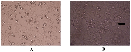
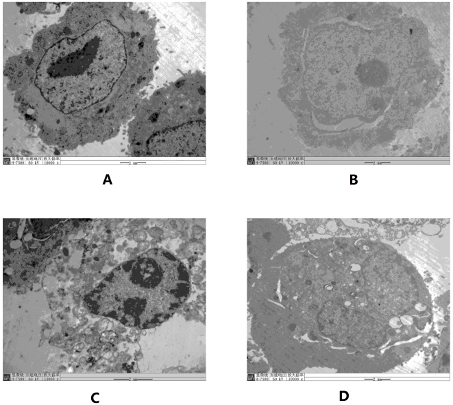
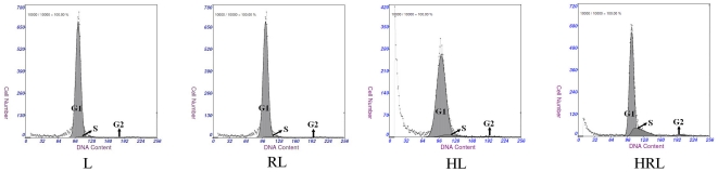
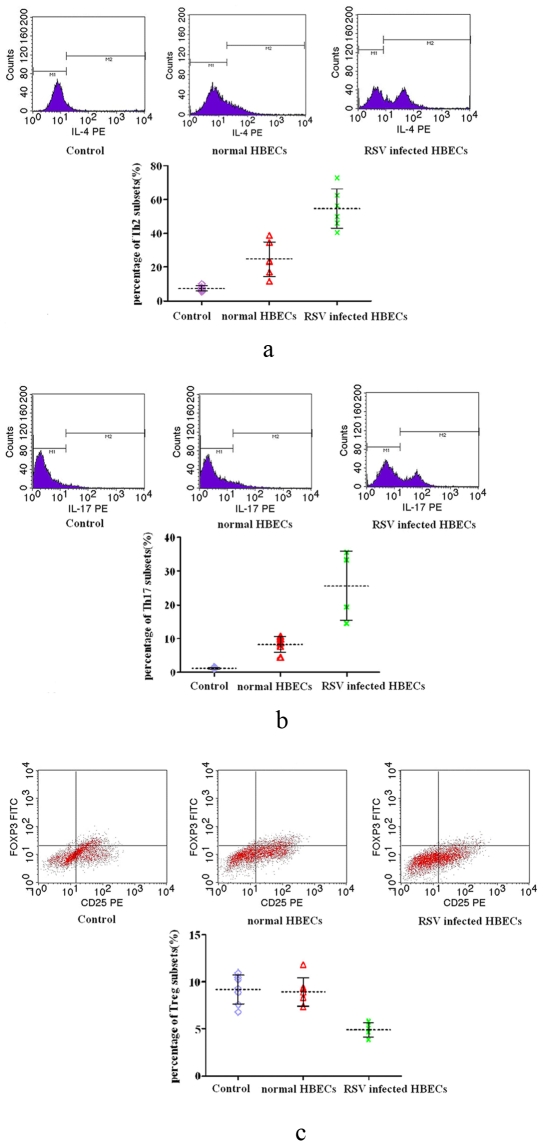
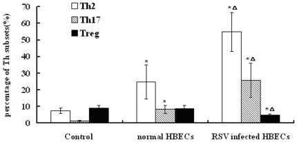
Respiratory syncytial virus (RSV) preferentially infects airway epithelial cells,which might be responsible for susceptibility to asthma; however, the underlying mechanism is not clear. This study determined the activation of lymphocytes and drift of helper T (Th) subsets induced by RSV-infected human bronchial epithelial cells (HBECs) in vitro. HBECs had prolonged infection with RSV, and lymphocytes isolated from human peripheral blood were co-cultured with RSV-infected HBECs. Four groups were established, as follows: lymphocytes (group L); lymphocytes infected with RSV (group RL); co-culture of lymphocytes with non-infected HBECs (group HL); and co-culture of lymphocytes with infected HBECs (group HRL). After co-culture with HBECs for 24 hours, lymphocytes were collected and the following were determined in the 4 groups: cell cycle status; apoptosis rate; and concentrations of IL-4, IFN-γ, and IL-17 in the supernatants. Cell cycle analysis for lymphocytes showed a significant increase in S phase cells, a decrease in G1 phase cells, and a higher apoptosis rate in group HRL compared with the other three groups. In group HRL, the levels of IL-4, IFN-γ, and IL-17 in supernatants were also higher than the other three groups. For further study, lymphocytes were individually treated with supernatants from non-infected and RSV-infected HBECs for 24 h. We showed that supernatants from RSV-infected HBECs induced the differentiation of Th2 and Th17 subsets, and suppressed the differentiation of Treg subsets. Our results showed that HBECs with prolonged RSV infection can induce lymphocyte proliferation and apoptosis, and enhance the release of cytokines by lymphocytes. Moreover, subset drift might be caused by RSV-infected HBECs.
© 2011 Qin et al.
Conflict of interest statement
Figures


Similar articles
-
[Th17/Treg imbalance mediated by IL-8 in RSV-infected bronchial epithelial cells].Zhong Nan Da Xue Xue Bao Yi Xue Ban. 2016 Apr;41(4):337-44. doi: 10.11817/j.issn.1672-7347.2016.04.001. Zhong Nan Da Xue Xue Bao Yi Xue Ban. 2016. PMID: 27241142 Chinese.
-
Leptin Is Oversecreted by Respiratory Syncytial Virus-Infected Bronchial Epithelial Cells and Regulates Th2 and Th17 Cell Differentiation.Int Arch Allergy Immunol. 2015;167(1):65-71. doi: 10.1159/000436966. Epub 2015 Jul 15. Int Arch Allergy Immunol. 2015. PMID: 26184438
-
Interleukin-23 facilitates Th1 and Th2 cell differentiation in vitro following respiratory syncytial virus infection.J Med Virol. 2015 Apr;87(4):708-15. doi: 10.1002/jmv.24126. Epub 2015 Feb 3. J Med Virol. 2015. PMID: 25648104
-
Interferon response and respiratory virus control are preserved in bronchial epithelial cells in asthma.J Allergy Clin Immunol. 2014 Dec;134(6):1402-1412.e7. doi: 10.1016/j.jaci.2014.07.013. Epub 2014 Sep 9. J Allergy Clin Immunol. 2014. PMID: 25216987 Free PMC article.
-
Respiratory syncytial virus (RSV) evades the human adaptive immune system by skewing the Th1/Th2 cytokine balance toward increased levels of Th2 cytokines and IgE, markers of allergy--a review.Virus Genes. 2006 Oct;33(2):235-52. doi: 10.1007/s11262-006-0064-x. Virus Genes. 2006. PMID: 16972040 Review.
Cited by
-
Modulation of host adaptive immunity by hRSV proteins.Virulence. 2014;5(7):740-51. doi: 10.4161/viru.32225. Virulence. 2014. PMID: 25513775 Free PMC article. Review.
-
New Insights Contributing to the Development of Effective Vaccines and Therapies to Reduce the Pathology Caused by hRSV.Int J Mol Sci. 2017 Aug 11;18(8):1753. doi: 10.3390/ijms18081753. Int J Mol Sci. 2017. PMID: 28800119 Free PMC article. Review.
-
Th17/IL-17 Axis Regulated by Airway Microbes Get Involved in the Development of Asthma.Curr Allergy Asthma Rep. 2020 Mar 14;20(4):11. doi: 10.1007/s11882-020-00903-x. Curr Allergy Asthma Rep. 2020. PMID: 32172346 Review.
-
The implication of infection with respiratory syncytial virus in pediatric recurrent wheezing and asthma: knowledge expanded post-COVID-19 era.Eur J Clin Microbiol Infect Dis. 2024 Mar;43(3):403-416. doi: 10.1007/s10096-023-04744-0. Epub 2023 Dec 28. Eur J Clin Microbiol Infect Dis. 2024. PMID: 38153660 Review.
-
Cytokine production and signaling pathways in respiratory virus infection.Front Microbiol. 2013 Sep 17;4:276. doi: 10.3389/fmicb.2013.00276. Front Microbiol. 2013. PMID: 24062733 Free PMC article. Review.
References
-
- Simoes EA. Respiratory syncytial virus infection. Lancet. 1999;354:847–852. - PubMed
-
- Hall CB, Long CE, Schnabel KC. Respiratory syncytial virus infections in previously healthy working adults. Clin Infect Dis. 2001;33:792–6. - PubMed
-
- Hall CB. Respiratory syncytial virus and parainfluenza virus. N Engl J Med. 2001;344:1917–28. - PubMed
-
- Busse WW. Mechanisms and advances in allergic diseases. J Allergy Clin Immunol. 2000;105:S593–8. - PubMed
Publication types
MeSH terms
LinkOut - more resources
Full Text Sources
