. 2011;6(12):e29112.
doi: 10.1371/journal.pone.0029112. Epub 2011 Dec 27.

Replication, gene expression and particle production by a consensus Merkel Cell Polyomavirus (MCPyV) genome

Affiliations

Replication, gene expression and particle production by a consensus Merkel Cell Polyomavirus (MCPyV) genome

Friederike Neumann et al. PLoS One. 2011.

Abstract

Merkel Cell Polyomavirus (MCPyV) genomes are clonally integrated in tumor tissues of approximately 85% of all Merkel cell carcinoma (MCC) cases, a highly aggressive tumor of the skin which predominantly afflicts elderly and immunosuppressed patients. All integrated viral genomes recovered from MCC tissue or MCC cell lines harbor signature mutations in the early gene transcript encoding for the large T-Antigen (LT-Ag). These mutations selectively abrogate the ability of LT-Ag to support viral replication while still maintaining its Rb-binding activity, suggesting a continuous requirement for LT-Ag mediated cell cycle deregulation during MCC pathogenesis. To gain a better understanding of MCPyV biology, in vitro MCPyV replication systems are required. We have generated a synthetic MCPyV genomic clone (MCVSyn) based on the consensus sequence of MCC-derived sequences deposited in the NCBI database. Here, we demonstrate that transfection of recircularized MCVSyn DNA into some human cell lines recapitulates efficient replication of the viral genome, early and late gene expression together with virus particle formation. However, serial transmission of infectious virus was not observed. This in vitro culturing system allows the study of viral replication and will facilitate the molecular dissection of important aspects of the MCPyV lifecycle.


Conflict of interest statement

Competing Interests: The authors have declared that no competing interests exist.

Figures

Figure 1
Figure 1. Alignment of MCVSyn and full length MCPyV sequences.
The illustration is based on a multiple sequence alignment using the Clustal W algorithm (see Table S3 for accession numbers used in the alignment) of MCVSyn and all full length MCPyV sequences deposited in the NCBI Database as of August 2011. Aligned genomes were compared to the consensus sequence (which is identical to MCVSyn as well as the isolates 17b, 18b and 20b). Nucleotide substitutions/mismatches relative to this sequence are shown as vertical black bars, whereas deletions are shown in red. Nucleotide insertions in a given sequence are shown as blue bars, and register as gaps in the backbone of the remaining genomes. Genomes that were isolated from MCC or MCC-derived cell lines are marked by an asterisk; the mutations which lead to the truncation of LT-Ag sequences in these genomes are likewise marked.
Figure 2
Figure 2. SV40 replication and gene expression in CV-1 cells transfected with SV40 DNA.
(A) 100 ng of intramolecular religated SV40 viral DNA was transfected in CV-1 cells and cells were lysed 12 h, 24 h, 36 h, 2d and 7d post transfection. Protein lysates were subsequently analyzed for SV40 LT-Ag (Pab419 antibody) and VP1 expression (α-VP1 polyclonal rabbit serum) by SDS-page and Western Blotting. Staining of actin was used to ensure that equal protein amounts were loaded per lane. (B) Low molecular weight DNA was isolated from SV40 DNA transfected CV-1 cells at the indicated time points by HIRT extraction, 1 µg DNA was DpnI and EcoRI digested; DNA was separated on an agarose gel and stained with EtBr (left panel), followed by southern blotting and detection of viral DNA using a 32PdCTP-labeled SV40 LT-Ag PCR fragment as a probe. The blot was exposed for 30 min. Numbers below the lanes correspond to the quantification of newly replicated DNA using a Fuji phosphoimager FLA7000 and MultiGauge software.
Figure 3
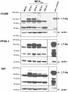
Figure 3. LT-Ag expression in human cell lines transfected with MCVSyn DNA.
5×10⁴ H1299, PSFK-1 or 293 cells were transfected with 100 ng re-circularized MCVSyn DNA or equivalent amounts of pUC18 DNA (Mock control). At the indicated time points, cells were lysed and analyzed by immunoblotting for MCPyV LT-Ag expression using the monoclonal LT-Ag antibody Cm2B4. Equal protein loading was confirmed by re-incubating the membrane with an anti-actin antibody. An LT-Ag expression control (pos. control; transient transfection with a CMV-promoter driven LT-Ag expression construct for 48 h) was loaded as an internal control.
Figure 4
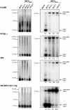
Figure 4. MCVSyn replication assays in H1299, PFSK-1 and 293 cells.
5 µg low molecular weight DNA isolated from cell cultures at the indicated time points post-transfection with MCVSyn DNA was digested with DpnI and EcoRI, separated on an agarose gel and stained with EtBr (left panels), then transferred via southern blot and probed with a radioactively labelled LT-Ag PCR fragment . The blot was exposed for 24 h and scanned using a Fuji phosphoimager FLA7000; MultiGauge software was used for quantification.
Figure 5
Figure 5. R17a replication in PFSK-1 cells.
2 µg low molecular weight DNA from PFSK-1 cells transfected with MCVSyn or R17a viral DNA was EcoRI and DpnI digested and separated on an agarose gel, followed by EtBr staining (left panel). The DNA was transferred via southern blotting and probed with a radioactively labelled LT-Ag PCR fragment (right panel). The blot was exposed for 24 h.
Figure 6
Figure 6. Real-time PCR to quantify early and late gene transcripts in cells transfected with MCVSyn.
RNA was isolated at the indicated time points after transfection, DNAse I digested and used for cDNA synthesis followed by real time PCR using a LT-Ag or VP1 specific primer set. Results were normalized against GAPDH transcript levels.
Figure 7
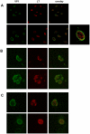
Figure 7. Subcellular localization of LT-Ag and VP1 protein in cells transfected with religated viral DNA.
(A) Double staining of CV-1 cells transfected with SV40 viral DNA. 4d p.t. the cells were fixed, and VP1 was detected with a polyclonal anti-VP1 antibody. LT-Ag was visualized with the monoclonal anti-LT antibody Pab419. Z-stack pictures were taken using confocal microscopy. Each picture represents an individual Z-stack. VP1 staining was observed primarily in speckles close to or at the nuclear membrane. LT-Ag staining was observed throughout the nucleoplasm with the nucleoli excluded. In some cells granular LT-Ag staining was observed. The panel on the lower right represents a 3× zoomed picture of a CV1 transfected cell with the two channels merged. Double staining of Merkel cell polyomavirus VP1 and LT-Ag in H1299 cells (B) and PFSK-1 cells (C) 4d p.t. reveals inner peripheral nuclear localization of MCVSyn VP1 protein. VP1 was visualized with a polyclonal anti-VP1 serum and anti-rabbit FITC, while LT-Ag was visualized with the monoclonal antibody Cm2B4 specifically recognizing MCPyV LT-Ag. 40 Z-stack pictures were taken scanning through the cells using a 63× magnification and 2fold zoom on a confocal microscope. The picture shown represents an individual image from the center of a Z-stack.
Figure 8
Figure 8. Density gradient centrifugation of SV40 and MCVSyn particles.
Optiprep™ gradient centrifugation was performed with cell lysates from CV1 (A) and H1299 (B) cells 4d after transfection with viral DNA. 15×250 µl fractions were collected (fraction 1 represents the fraction with the highest density and fraction 15 represents the lowest density fraction). (A) Left panel: Real time PCR of micrococcal nuclease treated fractions was performed using SV40 VP1 primer sequences. 20 µl of each gradient fraction was loaded on a 10% SDS-page followed immunoblotting using anti-VP1 serum. Right panel: Negative EM staining of SV40 particles identified in fraction 9. (B) Left panel: Real time PCR results of H1299 MCVSyn gradient fractions after micrococcal nuclease treatment using MCPyV VP1-specific primers. Right panel: Negative EM staining of particles identified in fractions 10 and 6.
Figure 9
Figure 9. Electron micrographs of viral particles in SV40-transfected CV-1 cells.
Images were prepared from CV-1 cultures at 4 days post transfection with SV40 DNA. (A) Accumulation of SV40 particles in an isolated nucleus of CV-1 cell cultures. (B) Particles close to the plasma membrane and projecting into the intercellular space between two CV-1 cells potentially represent a cell-to-cell transmission event of SV40 particles. Such events were rarely detected. (C) Large numbers of SV40 viral particles attached to membranous structures.
Figure 10
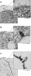
Figure 10. Electron micrographs of viral particles in MCVSyn-transfected PFSK-1 cells.
Images were prepared from PFSK-1 cultures at 8 days post transfection with MCVSyn DNA. (A and B) ∼40 µm electron dense particles were observed in approximately 1 out of 50 cells with the particles localizing in the nucleus close to membrane structures (additional particles in B that are located outside of the enlarged inset are marked by arrows). (C) Membrane-attached MCPyV particles, reminiscent of the structures observed in SV40 infected cells as shown in Figure 9C.

References

    1. Allander T, Andreasson K, Gupta S, Bjerkner A, Bogdanovic G, et al. Identification of a third human polyomavirus. J Virol. 2007;81:4130–4136. - PMC - PubMed
    1. Feng H, Shuda M, Chang Y, Moore PS. Clonal integration of a polyomavirus in human Merkel cell carcinoma. Science. 2008;319:1096–1100. - PMC - PubMed
    1. Gardner SD, Field AM, Coleman DV, Hulme B. New human papovavirus (B.K.) isolated from urine after renal transplantation. Lancet. 1971;1:1253–1257. - PubMed
    1. Gaynor AM, Nissen MD, Whiley DM, Mackay IM, Lambert SB, et al. Identification of a novel polyomavirus from patients with acute respiratory tract infections. PLoS Pathog. 2007;3:e64. - PMC - PubMed
    1. Padgett BL, Walker DL, ZuRhein GM, Eckroade RJ, Dessel BH. Cultivation of papova-like virus from human brain with progressive multifocal leucoencephalopathy. Lancet. 1971;1:1257–1260. - PubMed

Publication types