. 2011;6(12):e29313.
doi: 10.1371/journal.pone.0029313. Epub 2011 Dec 28.

The promigratory activity of the matricellular protein galectin-3 depends on the activation of PI-3 kinase

Affiliations

The promigratory activity of the matricellular protein galectin-3 depends on the activation of PI-3 kinase

Fabiana H M Melo et al. PLoS One. 2011.

Abstract

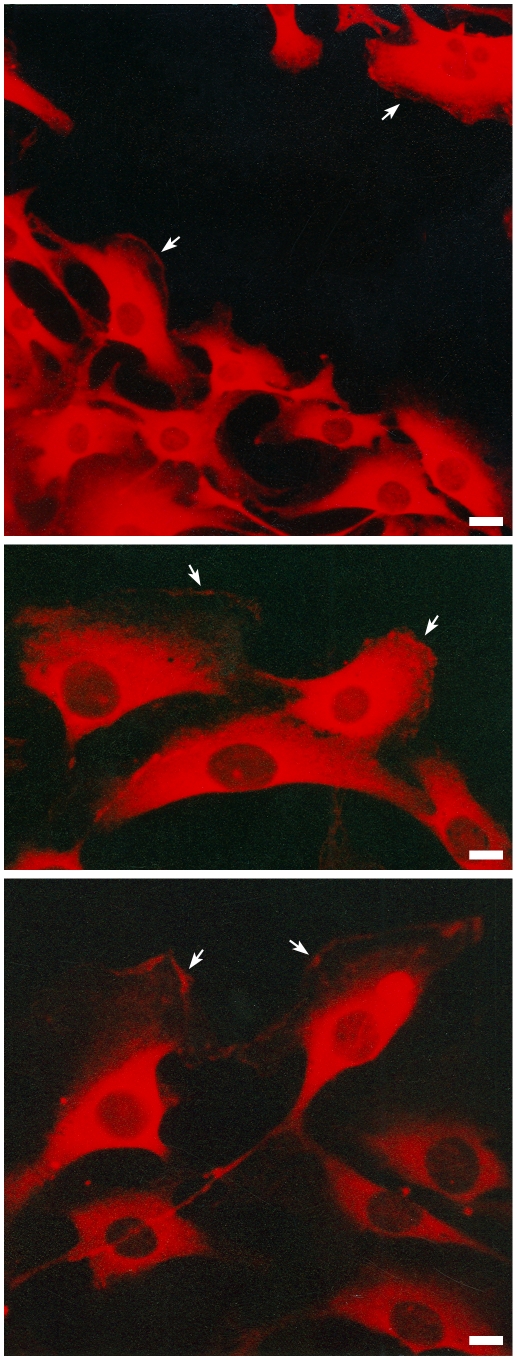
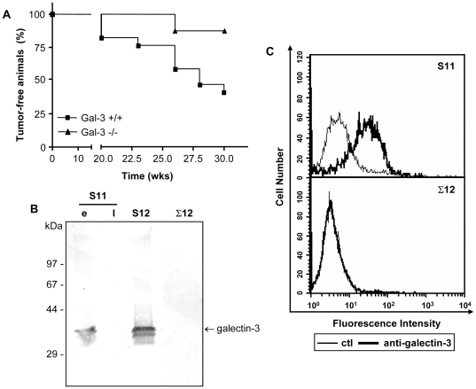
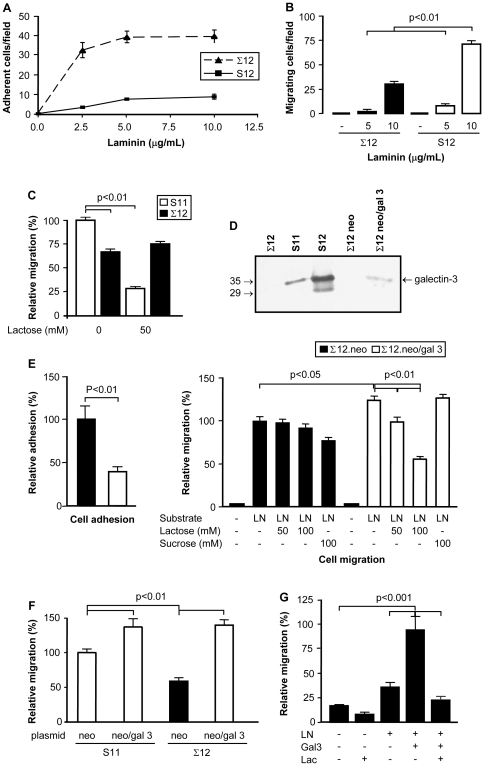
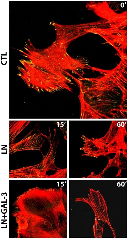
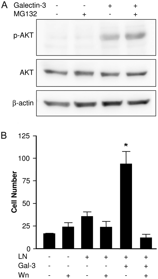
Expression of galectin-3 is associated with sarcoma progression, invasion and metastasis. Here we determined the role of extracellular galectin-3 on migration of sarcoma cells on laminin-111. Cell lines from methylcholanthrene-induced sarcomas from both wild type and galectin-3(-/-) mice were established. Despite the presence of similar levels of laminin-binding integrins on the cell surface, galectin-3(-/-) sarcoma cells were more adherent and less migratory than galectin-3(+/+) sarcoma cells on laminin-111. When galectin-3 was transiently expressed in galectin-3(-/-) sarcoma cells, it inhibited cell adhesion and stimulated the migratory response to laminin in a carbohydrate-dependent manner. Extracellular galectin-3 led to the recruitment of SHP-2 phosphatase to focal adhesion plaques, followed by a decrease in the amount of phosphorylated FAK and phospho-paxillin in the lamellipodia of migrating cells. The promigratory activity of extracellular galectin-3 was inhibitable by wortmannin, implicating the activation of a PI-3 kinase dependent pathway in the galectin-3 triggered disruption of adhesion plaques, leading to sarcoma cell migration on laminin-111.


Conflict of interest statement

Competing Interests: The authors have declared that no competing interests exist.

Figures

Figure 1
Figure 1. Galectin-3 is found on the lamellipodia of migrating fibroblasts.
CCR2 cells were subjected to the scrape assay. The cells were grown in coverslips to confluence, when a wound was made. The cells were maintained in culture for additional 24 hours, when it was possible to observe cells migrating into the wounded area. Panels show the distribution of galectin-3, which was detected with galectin-3 monoclonal antibody M3/38, followed by incubation with a rhodamine-conjugated secondary antibody. Galectin-3 is localized in the cytoplasm of the cells and also in leading edge of migrating cells as indicated by arrows.
Figure 2
Figure 2. Galectin-3−/− mice are more resistant to methylcholanthrene induced sarcomas development.
Wild type and galectin-3−/− mice were given two subcutaneous doses of 3-methylcholanthrene, a known chemical carcinogen. The development of tumors was followed weekly after the second dose. Analysis of the disease-free survival curves from both wild type (n = 23 animals) and galectin-3−/− mice (n = 17 animals) showed that wild type mice developed sarcomas faster than galectin-3−/− animals (A) (Log-rank test, p = 0.04). At necropsy, tumors were maintained in culture conditions, allowing for the establishment of sarcoma derived cell lines. Galectin-3 accumulation was analyzed in protein extracts from established cell lines from both wild type (S11 and S12) and galectin-3−/− mice (Σ12) by western blotting. S11 and S12 cells, but not Σ12 cells, expressed galectin-3. While S12 cells maintained high expression of galectin-3 in all passages analyzed, only early (e, passage number<15), but not late (l, passage number>15) passages of S11 expressed galectin-3. Note that part of the molecules produced were further processed rendering the lower molecular weight form of the lectin, which appears as a 30 kDa band (B). The expression of galectin-3 on S11 and Σ12 cell surface was analyzed by flow cytometry. S11 cells display at least part of its galectin-3 content on the cell surface (C).
Figure 3
Figure 3. The galectin-3−/− cell line is more adherent and less migratory on laminin-111 surfaces.
Σ12 cells were compared to S12 cells regarding their adhesive (panel A) and migratory capacity (panel B). For the adhesion assay (panel A), each point represents the mean of triplicates, SD is also represented. The migratory capacity of S12 and Σ12 cells on laminin-111 was evaluated using Transwell chambers (panel B). Migration assays were also performed comparing S11 and Σ12 cells, as described in B. In these assays, lactose was used to inhibit cell migration. S11 cell migration, but not Σ12 cell migration was inhibited by lactose. Migration of S11 cells without lactose inhibition was used as internal reference for determining relative migration (panel C). Σ12 cells were transiently transfected with either a plasmid containing the galectin-3 gene or a control plasmid. While no galectin-3 was found in Σ12 cells transfected with the control plasmid (neo) expression of galectin-3 was observed in neo/gal-3 transfected cells by western blotting. Extracts of S11 and S12 cells used were used as positive controls (panel D). Σ12 cells were rendered less adherent and more migratory upon transfection with pEF1-neo/gal-3. Relative adhesion was determined based on the adhesion of neo-transfected cells. Cell migration was evaluated in Transwell chambers, whose filters were coated with laminin-111. The transfected Σ12 cells were incubated in serum-free medium either in the absence or presence of the indicated concentrations of lactose or sucrose. Migration of the former cells towards laminin-111 was inhibited by lactose, but not by sucrose, indicating a role for galectin-3 in the modulation of cell migration in response to laminin-111 (panel E). S11 cells were transiently transfected with pEF1-neo or pEF1-neo/gal-3 and checked for their migratory response towards laminin-111 in Transwell chambers. A significant increase in the migratory response elicited by laminin-111 was observed in S11 cells overexpressing galectin-3. Migration of neo-transfected S11 cells was used as internal reference for determining relative migration (panel F). The migratory capacity of Σ12 cells was evaluated in absence or presence of extracellular galectin-3 (gal-3), laminin-111 (LN) and in the presence or absence of 100 mM lactose (Lac) for 24 hours using the scrape assay. Exogenous galectin-3 increased the migration of Σ12 cells when soluble LN was added, such increase was inhibited by lactose. Migration of Σ12 cells in the presence of both LN and galectin-3 was used as internal reference for determining relative migration (panel G). In all panels, results are representative of at least three independent assays. Means and SD are represented. White bars represent results from galectin-3 expressing cells; black bars represent results from galectin-3 null cells.
Figure 4
Figure 4. Extracellular galectin-3 (Gal-3) promotes the disassembly of stable focal adhesion plaques by decreasing the amount of phosphorylated FAK in the lamellipodia of migrating cells.
Σ12 cells were grown on coverslips and were subjected to the scrape assay either in the absence (ctl) or presence of laminin-111 (LN) or laminin-111 and 20 µg/mL galectin-3 (LN+gal-3) for 15 minutes. Cells were either fixed after 15 minutes after the migration stimulus. Confocal photomicrographs of typical fields are shown. Intracellular distribution of phosphorylated FAK (green) and organization of stress fibers (phalloidin staining, in red) are shown. Control cells showed mature adhesion plaques, as shown by staining of phosphorylated FAK. In the presence of galectin-3, there was a fast disassembly of the adhesion plaques indicated by the decrease of phosphorylated FAK in the lamellipodia.
Figure 5
Figure 5. Analysis of proteins enriched in the focal complexes in the absence (-) or presence (+) of extracellular galectin-3.
Σ12 cells were exposed to 10 µg/mL laminin and 20 µg/mL galectin-3 for 15 minutes and focal complexes were then prepared and analyzed regarding the presence of galectin-3 (A), SHP-2 (B) and phospho-paxillin (C) using Western blot analysis. (Panel A) Recruitment of galectin-3 to focal complexes was inhibited in the presence of lactose (lane at the right in panel A). Two bands were recognized by M3/38 monoclonal antibody in focal adhesion extracts; one which migrates with an apparent molecular weight of 30–35 kDa (Gal-3) and a second one which migrates as a 70 kDa-band (arrow). CE, in the left lane in panels A and B refer to Σ12 cellular extracts. (Panels B and C) Exposure of Σ12 cells to extracellular galectin-3 led to an increase in SHP-2 (panel B) and decrease of phosphorylated paxillin in the focal complexes (panel C). Prior inhibition of proteasome with lactacystin abrogated the decrease of phosphorylated paxillin in focal complexes. Representative blot of paxillin and phosphorylated paxillin are shown and the densitometric analysis of two independent experiments were performed, yielding similar results.
Figure 6
Figure 6. Galectin-3 increases Σ12 cell motility through a PI-3 kinase dependent pathway.
(A) Σ12 cells were exposed to 10 µg/mL laminin-111 either in the absence or presence of 20 µg/mL galectin-3 and of the proteasome inhibitor MG-132 for 15 minutes and phospho-AKT was analyzed using Western blot analysis. Extracellular galectin-3 leads to increase of phosphorylated AKT. Representative blot of AKT and phosphorylated AKT are shown and the densitometric analysis of two independent experiments were performed. (B) Σ12 cells were grown in coverslips and subjected to the scrape assay. Migration of Σ12 cells into the scratched area was measured after 24 hours by direct counting of DAPI-stained cells using a graticle projected onto micrographs collected using a fluorescence microscope. Cells were incubated or not in the presence of 10 µg/mL laminin-111 (LN), 20 µg/mL galectin-3 (Gal-3) and 1 mM wortmannin (Wn), as indicated. Bars represent mean of three independent assays done in triplicates, the SD is also represented. Analysis was done using one-way ANOVA, followed by the Bonferroni's t-test for multiple comparisons. There were no statistically significant differences among the groups treated in the absence of galectin-3. Exposure of Σ12 cells to galectin-3 and laminin-111 increased their motility about 2.5 fold over control conditions and over exposure of laminin-111 alone (p<0.001). The galectin-3 effect was inhibited by wortmannin (*, p<0.001), which did not affect significantly the migratory capacity of cells under the other experimental conditions.

Similar articles

Cited by

References

    1. Hanahan D, Weinberg RA. The hallmarks of cancer. Cell. 2000;100:57–70. - PubMed
    1. Hood JD, Cheresh DA. Role of integrins in cell invasion and migration. Nat Rev Cancer. 2002;2:91–100. - PubMed
    1. Aumailley M, Bruckner-Tuderman L, Carter WG, Deutzmann R, Edgar D, et al. A simplified laminin nomenclature. Matrix Biol. 2005;24:326–332. - PubMed
    1. Jasiulionis MG, Chammas R, Ventura AM, Travassos LR, Brentani RR. alpha6beta1-Integrin, a major cell surface carrier of beta1-6-branched oligosaccharides, mediates migration of EJ-ras-transformed fibroblasts on laminin-1 independently of its glycosylation state. Cancer Res. 1996;56:1682–1689. - PubMed
    1. Wehrle-Haller B, Imhof BA. Integrin-dependent pathologies. J Pathol. 2003;200:481–487. - PubMed

Publication types