. 2011;6(12):e29516.
doi: 10.1371/journal.pone.0029516. Epub 2011 Dec 28.

Polysialic acid is required for dopamine D2 receptor-mediated plasticity involving inhibitory circuits of the rat medial prefrontal cortex

Affiliations

Polysialic acid is required for dopamine D2 receptor-mediated plasticity involving inhibitory circuits of the rat medial prefrontal cortex

Esther Castillo-Gómez et al. PLoS One. 2011.

Abstract

Decreased expression of dopamine D2 receptors (D2R), dysfunction of inhibitory neurotransmission and impairments in the structure and connectivity of neurons in the medial prefrontal cortex (mPFC) are involved in the pathogenesis of schizophrenia and major depression, but the relationship between these changes remains unclear. The polysialylated form of the neural cell adhesion molecule (PSA-NCAM), a plasticity-related molecule, may serve as a link. This molecule is expressed in cortical interneurons and dopamine, via D2R, modulates its expression in parallel to that of proteins related to synapses and inhibitory neurotransmission, suggesting that D2R-targeted antipsychotics/antidepressants may act by affecting the plasticity of mPFC inhibitory circuits. To understand the role of PSA-NCAM in this plasticity, rats were chronically treated with a D2R agonist (PPHT) after cortical PSA depletion. PPHT-induced increases in GAD67 and synaptophysin (SYN) neuropil expression were blocked when PSA was previously removed, indicating a role for PSA-NCAM in this plasticity. The number of PSA-NCAM expressing interneuron somata also increased after PPHT treatment, but the percentages of these cells belonging to different interneuronal subpopulations did not change. Cortical pyramidal neurons did not express PSA-NCAM, but puncta co-expressing this molecule and parvalbumin could be found surrounding their somata. PPHT treatment increased the number of PSA-NCAM and parvalbumin expressing perisomatic puncta, but decreased the percentage of parvalbumin puncta that co-expressed SYN. PSA depletion did not block these effects on the perisomatic region, but increased further the number of parvalbumin expressing puncta and increased the percentage of puncta co-expressing SYN and parvalbumin, suggesting that the polysialylation of NCAM may regulate perisomatic inhibition of mPFC principal neurons. Summarizing, the present results indicate that dopamine acting on D2R influences structural plasticity of mPFC interneurons and point to PSA-NCAM as a key player in this remodeling.


Conflict of interest statement

Competing Interests: The authors have declared that no competing interests exist.

Figures

Figure 1
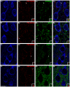
Figure 1. Confocal microscopic analysis of the neurochemical phenotype of PSA-NCAM immunoreactive puncta surrounding pyramidal cell somata in mPFC.
(A) PSA-NCAM-expressing puncta co-localizing with GAD67 in the perisomatic region of CaMKII-α expressing neurons. (B) PSA-NCAM/VGAT double-labeled puncta surrounding CaMKII-α immunoreactive neurons. (C) Lack of co-localization between perisomatic PSA-NCAM-expressing puncta and VGLUT1-expressing puncta. (D) Perisomatic puncta co-expressing PSA-NCAM and SYN. All the images in this figure are taken from single confocal planes. Scale bar: 10 µm. Insets in the images are 5× enlargements of puncta marked with arroheads.
Figure 2
Figure 2. Confocal microscopic analysis of the co-expression of PV, CR and CB in PSA-NCAM immunoreactive puncta surrounding pyramidal cell somata in mPFC.
(A) PSA-NCAM-expressing puncta co-expressing with PV in the perisomatic region of a neuron expressing CaMKII-α. (B) PSA-NCAM immunoreactive puncta co-expressing CR surrounding CaMKII-α expressing neurons. (C) Observe a CB-expressing puncta close to a PSA-NCAM expressing puncta in the perisomatic region of a pyramidal neuron. All the images in this figure are taken from single confocal planes. Scale bar: 10 µm. Insets in the images are 5× enlargements of puncta marked with arrowheads.
Figure 3
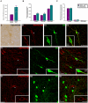
Figure 3. Quantification and confocal microscopic analysis of the neurochemical phenotype of PSA-NCAM expressing interneurons in mPFC after PPHT treatment.
(A) Graph showing statistically significant differences in the number of PSA-NCAM expressing somata from control group after unpaired Student's t-test; p<0.001 (***). (B) Graph representing the changes in the number of somata belonging to different interneuron subpopulations (defined by the expression of the calcium-binding proteins CB, CR and PV) after PPHT treatment [p<0.01 (**); p<0.001 (***) after unpaired Student's expression] (C) Graph showing the percentages of PSA-NCAM expressing interneuron somata belonging to different interneuron subpopulations. No statistically significant differences from control group were observed after unpaired Student's t-tests (p>0.05 in all comparisons). (D) Multipolar neuron expressing PSA-NCAM in dorsal Cingulate Cortex (Cg1) layer V observed under conventional light microscopy. (E) PSA-NCAM interneuron in Cg1 layer V co-expressing CB. (F) PSA-NCAM/CR double labeled interneuron in ventral Cingulate Cortex (Cg2) layer V. (G) Lack of co-localization between PSA-NCAM (G1) and PV (G2) expressing neurons in Cg1 layer III. E–G images are 2D projections of focal planes located 1 µm apart. Scale bar: 10 µm. Insets in the images are 2× enlarged views taken from single confocal planes of the areas marked with arrowheads.
Figure 4
Figure 4. Panoramic views of the rat mPFC cortex and graphs showing the expression of PSA-NCAM (a–e), SYN (f–j), GAD67 (k–o) and NCAM (p–t) in the mPFC neuropil after Endo-N or PPHT treatments and their combination.
Three sections per animal were examined under bright-field illumination, homogeneously lighted and digitalized using a CCD camera at 20× magnification. Grey levels were converted to optical densities (OD) using Image J software (NIH). Detailed description of this method of quantification can be found in material and methods S1. Note in panel c & d, the lack of PSA-NCAM expression in the cingulate cortex, but not in the striatum, demonstrating the effectiveness of Endo-N treatment. Asterisks in bars indicate statistically significant differences between groups (see graph legend) after univariate repeated measures ANOVA followed by multiple pair-wise comparisons with Bonferroni's correction; p<0.05 (*), p<0.01 (**), p<0.001 (***). Scale bar: 100 µm.
Figure 5
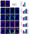
Figure 5. Confocal microscopic analysis of PSA-NCAM, GAD65/67, PV and SYN expressing puncta in the perisomatic region of mPFC pyramidal neurons after Endo-N or PPHT treatments and their combination.
Focal planes of pyramidal neuron somata (immunolabeled for CaMKII-α) showing the changes in the perisomatic density of PSA-NCAM (a–b), GAD65/67 (d–g), PV (i–l) and SYN (n–q) expressing puncta after the different treatments, as can be observed in graphs (c), (h), (m) and (r), respectively. (s–w) Focal planes and graph showing the changes on the percentage of PV expressing puncta co-expressing SYN after treatment. Asterisks in bars indicate statistically significant differences between groups (see graph legend) after unpaired Student's t-test (c) or one-way ANOVA (h, m, r, w) followed by Bonferroni's correction; p<0.05 (*), p<0.01 (**), p<0.001 (***).
Figure 6
Figure 6. Schematic drawing of the hypothetic effects of PPHT and/or Endo-N administration in the perisomatic region of pyramidal neurons and in the neuropil of the medial prefrontal cortex.
Perisomatic region: PPHT induces the appearance of new inhibitory contacts: some of them express PSA-NCAM (black asterisks), but some others may have already lost PSA-NCAM expression (red asterisks). Expression of PSA-NCAM may also be induced in preexisting inhibitory contacts (red arrow). It is possible that some of these PSA-NCAM expressing puncta were not functional synapses and this may be why we do not detect changes in the number of synaptophysin expressing puncta and there are decreases in the percentage of parvalbumin positive puncta that co-express synaptophysin. Endo-N depletes PSA-NCAM from the perisomatic region and promotes the maturation of contacts that previously expressed PSA-NCAM, inducing the expression of inhibitory markers (parvalbumin/GAD) and/or synaptophysin (black arrows), leading also to an increase of the percentage of parvalbumin puncta co-expressing synaptophysin. When PPHT is administered after Endo-N, most puncta remain stable, although some of them may continue with the maturation process initiated by PSA depletion and/or promoted by PPHT (green arrows). Neuropil: Note that the proportion of PSA-NCAM expressing puncta is more reduced than in the perisomatic region of pyramidal neurons and that many of these puncta do not correspond to synaptic boutons. PPHT induces the appearance of inhibitory puncta; some of them express PSA-NCAM (black asterisks) and some of them may have already lost its expression (red asterisks). Some of these newly generated structures may have matured faster than in the perisomatic region and have started to express synaptophysin. Endo-N administration may have little impact on neuropil because of the low proportion of PSA-NCAM expressing puncta in this region and because the nature of these puncta is different: most perisomatic puncta should correspond to presynaptic boutons contacting the pyramidal cell body, while in the neuropil many of these structures correspond to dendritic and axonal processes and not to synaptic contacts. The expression of inhibitory markers (parvalbumin/GAD) and/or synaptophysin would only be induced in some scarce puncta (black arrows). The administration of PPHT after Endo-N does not produce any effect, because the presence of PSA-NCAM may be absolutely necessary to induce the changes mediated by PPHT in this region.

Similar articles

Cited by

References

    1. Castren E. Is mood chemistry? Nat Rev Neurosci. 2005;6:241–246. - PubMed
    1. Lewis DA, Gonzalez-Burgos G. Neuroplasticity of neocortical circuits in schizophrenia. Neuropsychopharmacology. 2008;33:141–165. - PubMed
    1. Benes FM, Berretta S. GABAergic interneurons: implications for understanding schizophrenia and bipolar disorder. Neuropsychopharmacology. 2001;25:1–27. - PubMed
    1. Daskalakis ZJ, Fitzgerald PB, Christensen BK. The role of cortical inhibition in the pathophysiology and treatment of schizophrenia. Brain Res Rev. 2007;56:427–442. - PubMed
    1. Rajkowska G, O'Dwyer G, Teleki Z, Stockmeier CA, Miguel-Hidalgo JJ. GABAergic neurons immunoreactive for calcium binding proteins are reduced in the prefrontal cortex in major depression. Neuropsychopharmacology. 2007;32:471–482. - PMC - PubMed

Publication types