. 2011;6(12):e29539.
doi: 10.1371/journal.pone.0029539. Epub 2011 Dec 22.

The cytokine release inhibitory drug CRID3 targets ASC oligomerisation in the NLRP3 and AIM2 inflammasomes

Affiliations

The cytokine release inhibitory drug CRID3 targets ASC oligomerisation in the NLRP3 and AIM2 inflammasomes

Rebecca C Coll et al. PLoS One. 2011.

Erratum in

  • PLoS One. 2013;8(2). doi:10.1371/annotation/9f221489-155d-4978-a36d-30c51853e438
  • PLoS One. 2013;8(7). doi:10.1371/annotation/78d328b9-2c8c-4978-84b1-7e6a0b12ada1. Robertson, Avril [added]; Butler, Mark [added]; Cooper, Matthew [added]

Abstract

Background: The Inflammasomes are multi-protein complexes that regulate caspase-1 activation and the production of the pro-inflammatory cytokine IL-1β. Previous studies identified a class of diarylsulfonylurea containing compounds called Cytokine Release Inhibitory Drugs (CRIDs) that inhibited the post-translational processing of IL-1β. Further work identified Glutathione S-Transferase Omega 1 (GSTO1) as a possible target of these CRIDs. This study aimed to investigate the mechanism of the inhibitory activity of the CRID CP-456,773 (termed CRID3) in light of recent advances in the area of inflammasome activation, and to clarify the potential role of GSTO1 in the regulation of IL-1β production.

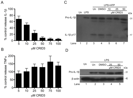
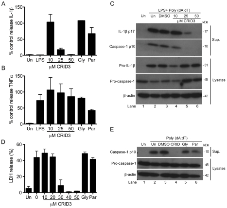
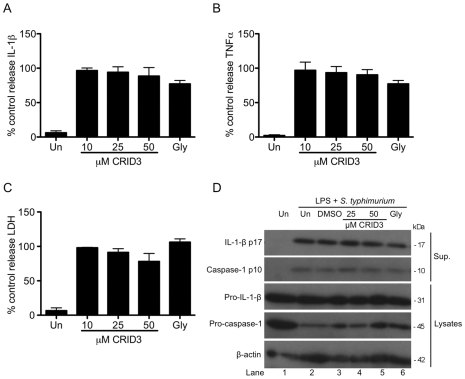
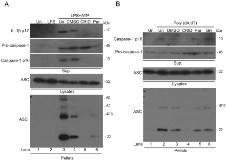
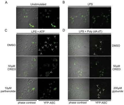
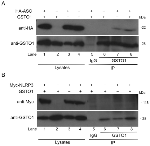
Methodology and results: In murine bone marrow derived macrophages, CRID3 inhibited IL-1β secretion and caspase 1 processing in response to stimulation of NLRP3 and AIM2 but not NLRC4. CRID3 also prevented AIM2 dependent pyroptosis in contrast to the NLRP3 inhibitors glyburide and parthenolide, which do not inhibit AIM2 activation. Confocal microscopy and Western blotting assays indicated that CRID3 inhibited the formation of ASC complexes or 'specks' in response to NLRP3 and AIM2 stimulation. Co-immunoprecipitation assays show that GSTO1 interacted with ASC.

Significance: These results identify CRID3 as a novel inhibitor of the NLRP3 and AIM2 inflammasomes and provide an insight into the mechanism of action of this small molecule. In addition GSTO1 may be a component of the inflammasome that is required for ASC complex formation.


Conflict of interest statement

Competing Interests: The authors have declared that no competing interests exist.

Figures

Figure 1
Figure 1. CRID3 inhibits LPS and ATP induced IL-1β processing.
Primary BMDM were stimulated with 10 ng/ml LPS for 3 h, treated with CRID3 (5–100 µM) or DMSO in serum free media for 30 min followed by addition of 5 mM ATP for 1 h. Supernatants were analysed by ELISA for IL -1β (A), TNFα (B) or by Western blotting for pro and mature IL-1β (C). (A) and (B) Cytokine level is expressed as a percentage of that released from control treated cells, which ranged across all experiments from 521–1988 pg/ml for IL-1β and 104–211 pg/ml for TNFα. Data are expressed as mean ± S.E.M of five independent experiments each carried out in triplicate. (C) A representative blot from one of three independent experiments is shown. (D) BMDM were treated with DMSO or CRID3 (25–50 µM) for 30 min and were then stimulated with 10 ng/ml LPS for 3 h. Protein samples were analysed by Western blotting using anti-IL-1β and anti-β-actin antibodies. A representative blot from one of two independent experiments is shown.
Figure 2
Figure 2. CRID3 inhibits the AIM2 inflammasome.
Primary BMDM were stimulated with 10 ng/ml LPS for 3 h, treated with CRID3 (10–50 µM), 200 µM glyburide, 10 µM parthenolide or DMSO control in serum free media for 1 h followed by transfection of 1 µg/ml Poly (dA:dT) for 4 h (A) and (B) or overnight (C). Supernatants were analysed by ELISA for IL -1β (A) and TNFα (B). (A) and (B) Cytokine level is expressed as a percentage of that released from control treated cells, which ranged across all experiments from 518–2347 pg/ml for IL-1β and 397–1136 pg/ml for TNFα. Data are expressed as mean ± S.E.M of four independent experiments each carried out in triplicate. In (C) concentrated supernatants and cell lysates were analysed by Western blotting using anti-IL-1β, anti-caspase-1 and anti-β-actin antibodies. These results are representative of three independent experiments. (D) and (E) Primary BMDM were treated with CRID3 (10–50 µM), 200 µM glyburide,10 µM parthenolide or DMSO in serum free media for 1 h followed transfection of 1 µg/ml Poly (dA:dT) for 6 h. Supernatants were analysed using an LDH cytotoxicity assay. The data shown represent mean % LDH release ± S.D. from triplicate determinations and are representative of three independent experiments. In (E) Concentrated supernatants and cell lysates were analysed by Western blotting using anti-caspase-1 and anti-β-actin antibodies. These results are representative of four independent experiments.
Figure 3
Figure 3. CRID3 and glyburide do not inhibit the NLRC4 inflammasome.
Primary BMDM were stimulated with 10 ng/ml LPS for 3 h, treated with CRID3 (10–50 µM), 200 µM glyburide or DMSO in serum free media for 30 min followed by infection with S. typhimurium (M.O.I 20) for 2 h. Supernatants were analysed by ELISA for IL -1β (A) and TNFα (B) production or by LDH cytotoxicity assay (C). (A) and (B) cytokine level is expressed as a percentage of that released from control treated cells, which ranged across all experiments from 534–3013 pg/ml for IL-1β and 1990–2746 pg/ml for TNFα. (A), (B) and (C) data are expressed as mean ± S.E.M of three independent experiments each carried out in triplicate. In (D) concentrated supernatants and cell lysates were analysed by Western blotting using anti-IL-1β, anti-caspase-1 and anti-β-actin antibodies. These results are representative of three independent experiments.
Figure 4
Figure 4. CRID3 prevents NLRP3 and AIM2 stimulated ASC oligomerisation.
(A) I-BMDM were stimulated with 10 ng/ml LPS for 3 h, were treated with 50 µM CRID3, 10 µM parthenolide, DMSO or were left untreated in serum free media for 30 min followed by addition of 5 mM ATP for 1 h. (B) Cells were treated with with 50 µM CRID3, 200 µM glyburide,10 µM parthenolide, DMSO or were left untreated in serum free media for 30 min followed by transfection with 1 µg/ml Poly (dA:dT) for 5 h. Concentrated supernatants and cell lysates were analysed by Western blotting using anti-IL-1β and/or anti-caspase-1 antibodies. The cell lysates were centrifuged at 330 x g for 10 min at 4°C. The pellets were washed and resuspended in PBS and then cross-linked by incubation with DSS for 30 min. The cross-linked pellets and cell lysates were analysed by Western blotting with anti-ASC and anti-caspase-1 antibodies. The data shown are representative of two (A) and three (B) independent experiments.
Figure 5
Figure 5. CRID3 inhibits NLRP3 and AIM2 stimulated ASC speck formation.
(A) ASC-YFP cells were left unstimulated, or were (B) stimulated with 10 ng/ml LPS for 3 h. (C) Cells were stimulated with 10 ng/ml LPS for 3 h, treated with 50 µM CRID3 10 µM parthenolide or DMSO in serum free media for 30 min followed by addition of 5 mM ATP for 1 h. In (D) cells were stimulated with 10 ng/ml LPS for 3 h, treated with 50 µM CRID3, 200 µM glyburide or DMSO in serum free media for 40 min followed by transfection of 1 µg/ml Poly (dA:dT) for 2 h. Cells were viewed by live cell imaging, at least four different images were taken of each dish of which a representative image is shown. These results are representative of three independent experiments.
Figure 6
Figure 6. ASC but not NLRP3 immunoprecipitates with GSTO1.
HEK-293T cells were transfected with HA-ASC, Myc-NLRP3, GSTO1 or empty vector plasmids as indicated. 48 hours post transfection cells were lysed in low stringency lysis buffer. 50 µl of lysate was kept for analysis while the remaining lysates were pre-cleared using 10 µl Protein A/G beads for 30 min. The lysates were then incubated with anti-GSTO1 antibody or rabbit IgG coupled to protein A/G beads for 3 h at 4°C. Whole cell lysates and immunoprecipitation samples were analysed by Western blotting using anti-Myc, anti-HA or anti-GSTO1 antibodies. The results presented are representative of two (B) and three (A) independent experiments.

References

    1. Bauernfeind F, Ablasser A, Bartok E, Kim S, Schmid-Burgk J, et al. Inflammasomes: current understanding and open questions. Cellular and molecular life sciences: CMLS. 2011;68:765–783. - PMC - PubMed
    1. Schroder K, Tschopp J. The inflammasomes. Cell. 2010;140:821–832. - PubMed
    1. Keller M, Ruegg A, Werner S, Beer HD. Active caspase-1 is a regulator of unconventional protein secretion. Cell. 2008;132:818–831. - PubMed
    1. Fernandes-Alnemri T, Wu J, Yu JW, Datta P, Miller B, et al. The pyroptosome: a supramolecular assembly of ASC dimers mediating inflammatory cell death via caspase-1 activation. Cell death and differentiation. 2007;14:1590–1604. - PMC - PubMed
    1. Agostini L, Martinon F, Burns K, McDermott MF, Hawkins PN, et al. NALP3 forms an IL-1beta-processing inflammasome with increased activity in Muckle-Wells autoinflammatory disorder. Immunity. 2004;20:319–325. - PubMed

Publication types