. 2011;6(12):e29599.
doi: 10.1371/journal.pone.0029599. Epub 2011 Dec 22.

Heregulin β-1 induces loss of cell-cell contact and enhances expression of MUC1 at the cell surface in HCC2998 and MKN45-1 cells

Affiliations

Heregulin β-1 induces loss of cell-cell contact and enhances expression of MUC1 at the cell surface in HCC2998 and MKN45-1 cells

Rintaro Okoshi et al. PLoS One. 2011.

Abstract

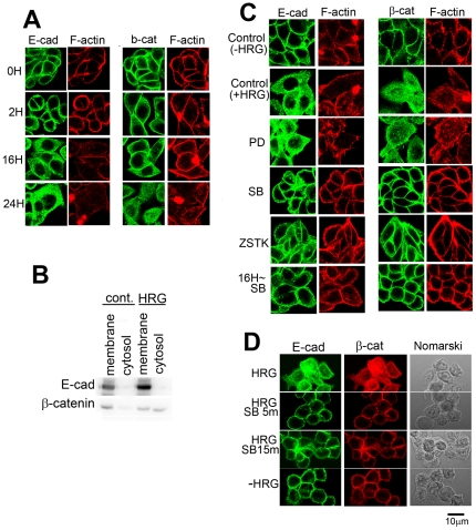
Signal transduction and cell responses after stimulation with heregulin β-1 (HRG) are examined in HCC2998 and MKN45-1 cells, which have been used for a model system to study the formation of signet ring carcinomas, one of poorly differentiated adenocarcinomas. HRG stimulation causes rounding of the cells, responding to HRG. The adherens junction, which is present in the control cells, is disrupted and cell-cell interaction is lost after stimulation. Inhibition of phosphatidylinositol (PI)-3 kinase or p38 MAP kinase blocked this reaction, which indicates that the PI-3 kinase-p38 MAP kinase pathway is required for this reaction. Inhibition of the p38 MAP kinase pathway resulted in immediate restoration of cell-cell interaction. This result indicates that signaling for adherent molecules is strictly regulated by growth factor signaling. Expression of MUC1 at the cell surface is also observed and found to be expressed only after HRG stimulation. The total amount of MUC1 remains unchanged, suggesting that this amount is not due to induction of gene expression but to translocation of MUC1 from the inner membrane to the plasma membrane. This reaction is independent of the cytohesin pathway but dependent on PI-3 kinase activity. In addition to these reactions, HRG stimulates cell growth of both HCC2998 and MKN45-1 cells, depending on the ERK pathway given that the MEK inhibitor abolishes this effect. Therefore, HRG induces various reactions in HCC2998 and MKN45-1 cells by different pathways. These reactions are all related to characteristics of tumors, which implicates that HRG signaling can contribute to the formation of tumors.


Conflict of interest statement

Competing Interests: The authors have declared that no competing interests exist.

Figures

Figure 1
Figure 1. Reaction of HCC2998 cells to HRG stimulation.
(A) HCC2998 cells were treated with HRG (100 ng/ml) for the indicated time. Cells were observed under the microscope. “∼16 h SB” indicates addition of SB202190 (1 µM) to the culture after incubation with HRG for 16 h. (B) HCC2998 cells were treated with HRG (100 ng/ml) for the indicated time. Protein levels and their phosphorylation were analyzed by Western blotting. (C) HCC2998 cells were treated with HRG in the presence of PD98059 (PD), SB202190 (SB), or ZSTK474 (ZSTK) for 24 h. Cells were observed under the microscope.
Figure 2
Figure 2. Reaction of MKN45-1 cells to HRG stimulation.
(A) MKN45-1 cells were treated with HRG (100 ng/ml) for the indicated time. Cells were observed under the microscope. (B) HCC2998 cells were treated with HRG (100 ng/ml) for the indicated time. Protein levels and their phosphorylation were analyzed by Western blotting. (C) HCC2998 cells were treated with HRG in the presence of PD98059 (PD), SB202190 (SB), or ZSTK474 (ZSTK) for 24 h. Cells were observed under the microscope.
Figure 3
Figure 3. E-cadherin and β-catenin in HCC2998 cells stimulated with HRG.
(A) HCC2998 cells were stimulated with HRG for the indicated time. E-cadherin or β-catenin was co-stained with F-actin. (B) HCC2998 cells were treated with HRG for 24 h. Cytoplasmic fraction of the cells was further fractionated into the membrane and the cytosolic fractions. The membrane fraction was dissolved in the same volume of the sample buffer used for electrophoresis of the cytosolic fraction. The resulting samples were analyzed for the levels of E-cadherin and β-catenin by Western blotting. (C) Effect of inhibitors for various signaling molecules (PD98059, 10 µM; SB202190, 1 µM; ZSTK474, 1 µM) on HCC2998 cells treated with HRG for 24 h. E-cadherin or β-catenin was co-stained with F-actin. “16h∼ SB” indicates addition of SB202190 (1 µM) to the culture after incubation with HRG for 16 h and further incubation for 8 h. Sections at about the center of the cells are shown. (D) Cells were treated with HRG for 22 h. Then 1 µM SB202190 was added for the indicated time. Cell morphology and localization of E-cadherin was monitored by immunostaining. Sections of the cells at about the middle parts are shown.
Figure 4
Figure 4. E-cadherin and β-catenin in MKN45-1 cells stimulated with HRG.
(A) MKN45-1 cells were stimulated with HRG for the indicated time. E-cadherin or β-catenin was co-stained with F-actin. Effect of inhibitors for various signaling molecules (PD98059, 10 µM; SB202190, 1 µM; ZSTK474, 1 µM) on MKN45-1 cells treated with HRG for 24 h. (B) Cells were treated with HRG for 22 h. Then 1 µM SB202190 was added for the indicated time. Cell morphology and localization of E-cadherin were monitored by immunostaining. Sections of the cells at about the middle parts are shown.
Figure 5
Figure 5. Expression of MUC1 at the cell surface after stimulation with HRG.
(A) HCC2998 cells were stimulated with HRG for the indicated time. A second dose of HRG was given after incubation for 24 h for the 2-day cultures. Muc1, with or without permeabilization, was stained with anti-MUC1 antibody. On the right sides, Nomarski view of the same cells are shown. (B) HCC2998 cells were stimulated with HRG for the indicated time. Total MUC1 levels and MUC1 at the cell surface (biotinylation) were analyzed by Western blotting. Detecting MUC1 expressed at the cell surface required 50 times more protein than was used for simple Western blotting. (C) Effect of inhibitors for various signaling molecules on MUC1 expression at the cell surface was examined. HCC2998 cells were cultured for 2 days in the presence of HRG. (D) HCC2998 cells bearing inducible genes for dominant active PI-3 kinase (BD110) and MKK6 were used , . They were infected with adenovirus bearing Cre recombinase to induce the gene. Adenovirus bearing LacZ was used as a control. (E) Experiments similar to C were done using SecinH3 (20 µg/ml).
Figure 6
Figure 6. Expression of MUC1 at the cell surface after stimulation with HRG.
(A) MKN45-1 cells were stimulated with HRG for the indicated time. A second dose of HRG was given after incubation for 24 h for the 2-day cultures. Muc1, with or without permeabilization, was stained with anti-MUC1 antibody. On the right sides, Nomarski views of the same cells are shown. (B) Effect of inhibitors for various signaling molecules on MUC1 expression at the cell surface was examined. MKN45-1 cells were cultured for 2 days in the presence of HRG.
Figure 7
Figure 7. Flow cytometry of HCC2998 and MKN45-1 cells.
HCC2998 and MKN45-1 cells treated with or without HRG for 2 days. A second dose of HRG was given 24 h after the first dose. Cells were fixed, dispersed, and stained with an anti-MUC1 antibody, followed by the second antibody conjugated with Alexa488. The resulting cells were subjected to flow cytometry.
Figure 8
Figure 8. Effect of inhibitors for signaling molecules on growth of HCC2998 and MKN45-1 cells.
(A and B) HCC2998 cells were plated in 3.5 cm dishes at a cell density of 1×10⁴ and stimulated with HRG every day in the presence or absence of drugs. Cells in the dishes were counted. (C and D) Similar experiments were done with MKN45-1 cells. Experiments were done three times and plotted on the graph. The standard deviations for each point were less than 4%. Control: without HRG treatment.
Figure 9
Figure 9. Signal transduction of HRG in HCC2998 cells.
HRG induces various cell responses through different signaling pathways.

Similar articles

Cited by

References

    1. Kanakry CG, Li Z, Nakai Y, Sei Y, Weinberger DR. Neuregulin-1 regulates cell adhesion via an ErbB2/phosphoinositide-3 kinase/Akt-dependent pathway: potential implications for schizophrenia and cancer. PLoS One. 2007;2:e1369. - PMC - PubMed
    1. Yokota Y, Kim WY, Chen Y, Wang X, Stanco A, et al. The adenomatous polyposis coli protein is an essential regulator of radial glial polarity and construction of the cerebral cortex. Neuron. 2009;61:42–56. - PMC - PubMed
    1. Vermeer PD, Einwalter LA, Moninger TO, Rokhlina T, Kern JA, et al. Segregation of receptor and ligand regulates activation of epithelial growth factor receptor. Nature. 2003;422:322–326. - PubMed
    1. Falls DL. Neuregulins: functions, forms, and signaling strategies. Exp Cell Res. 2003;284:14–30. - PubMed
    1. Jones FE, Jerry DJ, Guarino BC, Andrews GC, Stern DF. Heregulin induces in vivo proliferation and differentiation of mammary epithelium into secretory lobuloalveoli. Cell Growth Differ. 1996;7:1031–1038. - PubMed

Publication types