. 2012 Mar 15;442(3):681-92.
doi: 10.1042/BJ20111530.

Identification of autophosphorylation sites in eukaryotic elongation factor-2 kinase

Affiliations

Identification of autophosphorylation sites in eukaryotic elongation factor-2 kinase

Sébastien Pyr Dit Ruys et al. Biochem J. .

Erratum in

  • Biochem J. 2012 Apr 1;443(1):328

Abstract

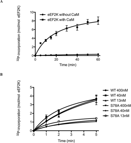
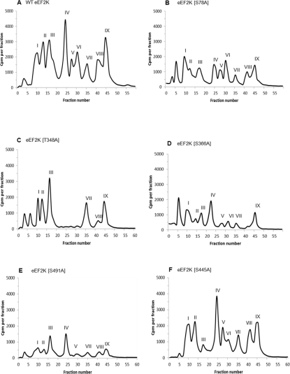
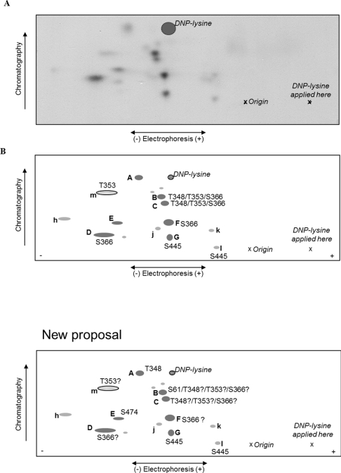
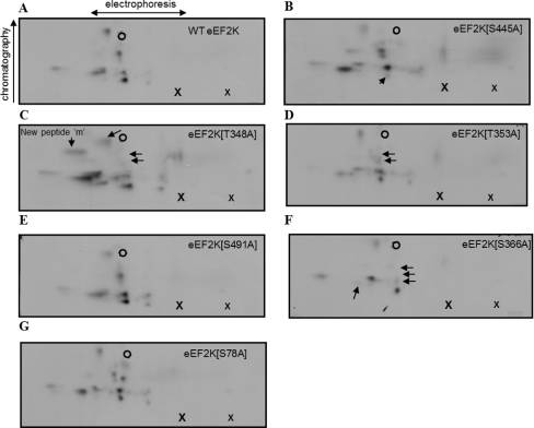
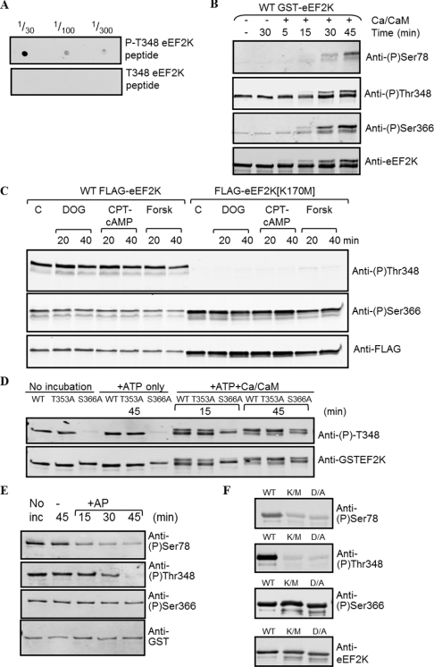
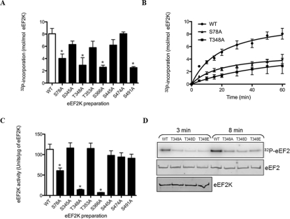
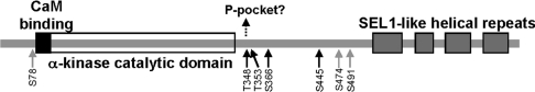
eEF2K [eEF2 (eukaryotic elongation factor 2) kinase] phosphorylates and inactivates the translation elongation factor eEF2. eEF2K is not a member of the main eukaryotic protein kinase superfamily, but instead belongs to a small group of so-called α-kinases. The activity of eEF2K is normally dependent upon Ca(2+) and calmodulin. eEF2K has previously been shown to undergo autophosphorylation, the stoichiometry of which suggested the existence of multiple sites. In the present study we have identified several autophosphorylation sites, including Thr(348), Thr(353), Ser(366) and Ser(445), all of which are highly conserved among vertebrate eEF2Ks. We also identified a number of other sites, including Ser(78), a known site of phosphorylation, and others, some of which are less well conserved. None of the sites lies in the catalytic domain, but three affect eEF2K activity. Mutation of Ser(78), Thr(348) and Ser(366) to a non-phosphorylatable alanine residue decreased eEF2K activity. Phosphorylation of Thr(348) was detected by immunoblotting after transfecting wild-type eEF2K into HEK (human embryonic kidney)-293 cells, but not after transfection with a kinase-inactive construct, confirming that this is indeed a site of autophosphorylation. Thr(348) appears to be constitutively autophosphorylated in vitro. Interestingly, other recent data suggest that the corresponding residue in other α-kinases is also autophosphorylated and contributes to the activation of these enzymes [Crawley, Gharaei, Ye, Yang, Raveh, London, Schueler-Furman, Jia and Cote (2011) J. Biol. Chem. 286, 2607-2616]. Ser(366) phosphorylation was also detected in intact cells, but was still observed in the kinase-inactive construct, demonstrating that this site is phosphorylated not only autocatalytically but also in trans by other kinases.


Figures

Figure 1
Figure 1. Recombinant eEF2K undergoes Ca2+/CaM-dependent autophosphorylation
(A) GST–eEF2K was incubated with [γ-32P]ATP in the presence or absence of Ca2+/CaM as described in the Experimental section. At the indicated times, samples were analysed by SDS/PAGE for measurements of 32P incorporation. The values are means±S.E.M. (n=3). (B) Autophosphorylation was measured as in (A) with the indicated concentrations of wild-type (WT) eEF2K and eEF2K[S78A] mutant.
Figure 2
Figure 2. HPLC profiles of 32P-autophosphorylated wild-type and mutant eEF2K preparations following trypsin digestion
Tryptic phosphopeptides from autophosphorylated eEF2K were separated by reverse-phase HPLC as described in the Experimental section. The major radiolabelled peaks are indicated by roman numerals. Phosphorylation sites detected in the peaks are summarized in Table 1 and discussed in the text. HPLC profiles are shown for wild-type (WT) eEF2K (A) and the eEF2K[S78A] (B), eEF2K[T348A] (C), eEF2K[S366A] (D), eEF2K[S491A] (E) and eEF2K[S445A] (F) mutants.
Figure 3
Figure 3. 2D peptide maps from autophosphorylated wild-type eEF2K
Wild-type eEF2K was allowed to undergo autophosphorylation in the presence of Ca2+/CaM and then subjected to tryptic digestion. Phosphopeptides were resolved by electrophoresis and chromatography (polarity and directions are indicated). Also shown are the position of the origin, where the peptide samples were applied, “X”, and the final migration position of the DNP–lysine marker (cross-hatched circle). (A) Representative map of wild-type eEF2K; (B) schematic summary of peptides; lettering in capitals for major peptides (shown in dark grey) and in lower case for minor ones (light grey). The peptide shown by the dotted oval (‘m’) was only observed on maps from the eEF2K[T348A] mutant.
Figure 4
Figure 4. 2D peptide maps from autophosphorylated wild-type eEF2K and mutants
Wild-type or mutant eEF2K was allowed to undergo autophosphorylation in the presence of Ca2+/CaM and then subjected to tryptic digestion. Phosphopeptides were resolved by electrophoresis and chromatography (polarity and directions are indicated). The positions where the sample (larger ‘X’) and the DNP–lysine (smaller “x”) were applied, and the final migration position of the DNP–lysine marker (open circle) are also shown. The maps are derived from wild-type eEF2K (A), eEF2K[S445A] (B), eEF2K[T348A] (C), eEF2K[T353A] (D), eEF2K[S491A] (E), eEF2K[S366A] (F) and eEF2K[S78A] (G). The arrows highlight spots that were lost or that decreased in intensity compared with the map from wild-type eEF2K. All peptide maps were run at least twice to verify their reproducibility, and similar levels of radioactivity were applied.
Figure 5
Figure 5. Use of phosphospecific antibodies to study the autophosphorylation of eEF2K in vitro and in intact cells
(A) Dot blot to establish whether the anti-phospho-Thr348 antibody is truly phosphospecific. A total of 1 μl of the indicated dilutions of the phosphorylated and non-phosphorylated versions of the corresponding peptide (10 mg/ml) were applied to the membrane, which was developed as for Western blot analysis. (B) Recombinant GST–eEF2K expressed in E. coli was incubated without or with Ca2+/CaM, as indicated, and non-radioactive ATP for the times shown. Samples were then analysed by SDS/PAGE followed by Western blot analysis with the indicated antibodies specific for phospho (P)-Ser78, phospho-Ser366 or phospho-Thr348, and anti-eEF2K antibody as a loading control. (C) Results for the phosphorylation of Thr348 and Ser366 in vivo. HEK-293 cells were transfected with a vector encoding either wild-type (WT) eEF2K or the kinase-defective eEF2K[K170M] mutant. Cells were treated for the indicated times with 2-deoxyglucose (DOG; 10 mM; after transfer to medium containing 5 mM glucose), chlorophenylthio-cAMP (CPT-cAMP; 0.5 mM) or forskolin (Forsk; 10 μM); the latter two were used in the presence of isobutylmethylxanthine (1 mM). Cell lysates were analysed by SDS/PAGE and immunoblotting using the indicated antibodies. C, control (untreated) cells. (D) Recombinant GST–eEF2K was incubated, where noted, with non-radioactive MgATP and, in some cases, Ca2+/CaM for the indicated times. Samples were then denatured and analysed by SDS/PAGE and Western blotting using the indicated antibodies. (E) Recombinant GST–eEF2K was either analysed directly (‘No inc’) or incubated with alkaline phosphatase (+AP) for the times shown. Samples were analysed by SDS/PAGE and Western blotting using the indicated antibodies. (F) Wild-type eEF2K or two kinase-deficient mutants (K/M, eEF2K[K170M]; D/A, eEF2K[D274A]) were analysed by SDS/PAGE and Western blotting using the indicated antibodies.
Figure 6
Figure 6. Autophosphorylation of wild-type and mutant eEF2K preparations and effects on eEF2K activity
(A) The histogram shows stoichiometries of autophosphorylation of the wild-type (WT) and mutant eEF2K preparations calculated after 60 min of incubation and, in (B), a time-course of phosphorylation of wild-type against the selected mutants is shown. The results are means±S.E.M., n=3. (C) eEF2K wild-type and mutant preparations were allowed to autophosphorylate in the presence of non-radioactive ATP as described in the Experimental section. After 60 min of incubation, aliquots were taken for eEF2K assay with MH-1 as substrate. Activities calculated from initial rates of 32P-incorporation are shown in the histogram. Values are means±S.E.M., n=3 and *P<0.001 compared with the wild-type. (D) The wild-type and indicated mutant eEF2K preparations were incubated with [γ-32P]ATP for autophosphorylation as described in the Experimental section. At the indicated times, samples were taken for SDS/PAGE and autoradiography. The bottom panel shows a Western blot for eEF2K to confirm that equal amounts of wild-type and mutant enzyme were used.
Figure 7
Figure 7. Location of autophosphorylation sites within the domain structure of eEF2K
The layout of the eEF2K polypeptide is depicted schematically (and only roughly to scale). The major sites of autophosphorylation identified in the present study are indicated (black arrows), with minor ones shown by grey arrows. The dotted arrow indicates that phosphorylated Thr348 may dock with the phosphate (P-) pocket in the C-terminal lobe of the catalytic domain to promote eEF2K activity, similar to Thr825 in MHCKA [25]. See the Discussion for further information.

References

    1. Ovchinnikov L. P., Motuz L. P., Natapov P. G., Averbuch L. J., Wettenhall R. E., Szyszka R., Kramer G., Hardesty B. Three phosphorylation sites in elongation factor 2. FEBS Lett. 1990;275:209–212. - PubMed
    1. Price N. T., Redpath N. T., Severinov K. V., Campbell D. G., Russell J. M., Proud C. G. Identification of the phosphorylation sites in elongation factor-2 from rabbit reticulocytes. FEBS Lett. 1991;282:253–258. - PubMed
    1. Carlberg U., Nilsson A., Nygard O. Functional properties of phosphorylated elongation factor 2. Eur. J. Biochem. 1990;191:639–645. - PubMed
    1. Ryazanov A. G., Davydova E. K. Mechanism of elongation factor 2 (EF-2) inactivation upon phosphorylation. Phosphorylated EF-2 is unable to catalyze translocation. FEBS Lett. 1989;251:187–190. - PubMed
    1. Drennan D., Ryazanov A. G. α-Kinases: analysis of the family and comparison with conventional protein kinases. Prog. Biophys. Mol. Biol. 2004;85:1–32. - PubMed

Publication types