. 2012 Jan 5;19(1):3.
doi: 10.1186/1423-0127-19-3.

Effects of phorbol myristate acetate and sivelestat on the lung injury caused by fat embolism in isolated lungs

Affiliations

Effects of phorbol myristate acetate and sivelestat on the lung injury caused by fat embolism in isolated lungs

Chia-Chih Lin et al. J Biomed Sci. .

Abstract

Background: Fat embolism syndrome (FES) associated with acute lung injury (ALI) is a clinical condition following long bone fracture. We have reported 14 victims due to ALI with FES. Our laboratory has developed an animal model that produced fat emboli (FE). The major purpose of this study was to test whether neutrophil activation with phorbol myristate acetate (PMA) and inhibition with sivelestat (SVT) exert protection on the lung.

Methods: The lungs of Sprague-Dawley rats were isolated and perfused. FE was produced by addition of corn oil micelles into the lung perfusate. PMA and SVT were given simultaneously with FE. Parameters such as lung weight/body weight ratio, LW gain, exhaled nitric oxide (NO), protein concentration in bronchoalveolar lavage relating to ALI were measured. The neutrophil elastase (NE), myeloperoxidase, malondialdehyde and phopholipase A₂ activity were determined. We also measured the nitrate/nitrite, methyl guanidine (MG), and cytokines. Pulmonary arterial pressure and microvascular permeability were assessed. Lung pathology was examined and scored. The inducible and endothelial NO synthase (iNOS and eNOS) were detected.

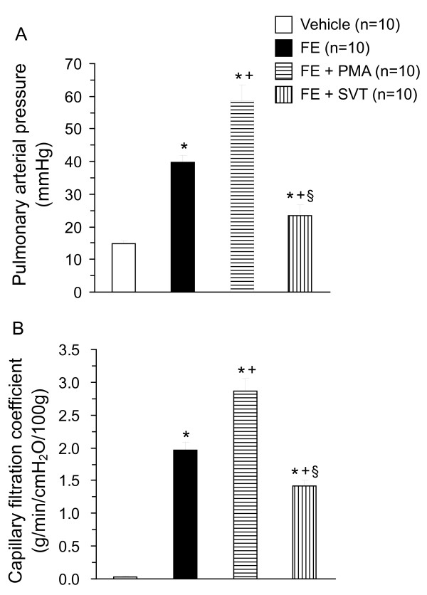
Results: FE caused ALI and increased biochemical factors. The challenge also resulted in pulmonary hypertension and increased microvascular permeability. The NE appeared to be the first to reach its peak at 1 hr, followed by other factors. Coadministration with PMA exacerbated the FE-induced changes, while SVT attenuated the effects of FE.

Conclusions: The FE-induced lung changes were enhanced by PMA, while SVT had the opposite effect. Sivelestat, a neutrophil inhibitor may be a therapeutic choice for patients with acute respiratory distress syndrome (ARDS) following fat embolism.


Figures

Figure 1
Figure 1
Lung weight/body weight (LW/BW) ratio (A), LW gain (LWG) (B), exhaled NO (C) and PCBAL (D) in isolated perfused lung following fat embolism (FE), FE + phorbol myristate acetate (PMA) and FE + sivelestat (SVT). *p < 0.05 vs. vehicle; +p < 0.05 FE + PMA vs. FE; and §p < 0.05 FE + SVT vs. FE.
Figure 2
Figure 2
Increases in pulmonary arterial pressure (A) and microvascular permeability (B) in isolated lung caused by FE. Cotreatment with PMA enhanced the pulmonary hypertension and capillary filtration, while FE with SVT attenuated the changes in pulmonary arterial pressure and capillary permeability. *p < 0.05 vs. vehicle; +p < 0.05 FE + PMA vs. FE; and §p < 0.05 FE + SVT vs. FE.
Figure 3
Figure 3
Histopathology of the lung tissue in vehicle (A), FE (B), FE + PMA (C) and FE + SVT (D) groups. FE caused severe alveolar edema and hemorrhage with inflammatory cell infiltration. Coadministration with PMA enhanced, but SVT diminished the lung pathology.
Figure 4
Figure 4
Time course of changes in neutrophil elastase (A), myeloperoxidase (B), malondialdehyde (C) and phospholipase A2 (D) in lung perfusate of vehicle, FE, FE + PMA and FE + SVT groups. *p < 0.05 vs. vehicle; +p < 0.05 FE + PMA vs. FE; and §p < 0.05 FE + SVT vs. FE.
Figure 5
Figure 5
Biochemical changes of nitrate/nitrite (A), methyl guanidine (B), tumor necrosis factorα (C) and interleukin-1β (D) in various groups. FE significantly elevated these biochemical factors. Cotreatment with PMA exacerbated these changes, while FE with SVT mitigated the changes in these mediators. *p < 0.05 vs. vehicle; +p < 0.05 FE + PMA vs. FE; and §p < 0.05 FE + SVT vs. FE.
Figure 6
Figure 6
Expression of inducible NO synthase (iNOS) and endothelial NO synthase (eNOS) mRNA by reverse transcriptase chain reaction in various groups (A). The iNOS/GAPDH and eNOS/GAPDH ratios (B) were increased by FE (*p < 0.05). Coadministration with PMA increased the iNOS and eNOS expression (+p < 0.05), while SVT attenuated the mRNA expression of iNOS and eNOS (§p < 0.05).

Similar articles

Cited by

References

    1. Hsu YH, Kao SJ, Lee RP, Chen HI. Acute pulmonary oedema: rare causes and possible mechanisms. Clin Sci (Lond) 2003;104:259–264. doi: 10.1042/CS20020166. - DOI - PubMed
    1. Karagiorga G, Nakos G, Galiatsou E, Lekka ME. Biochemical parameters of bronchoalveolar lavage fluid in fat embolism. Intensive Care Med. 2006;32:116–123. doi: 10.1007/s00134-005-2868-x. - DOI - PubMed
    1. Kao SJ, Yeh DY, Chen HI. Clinical and pathological features of fat embolism with acute respiratory distress syndrome. Clin Sci (Lond) 2007;113:279–285. doi: 10.1042/CS20070011. - DOI - PubMed
    1. White T, Petrisor BA, Bhandari M. Prevention of fat embolism syndrome. Injury. 2006;37:S59–S67. - PubMed
    1. Liu DD, Hsieh NK, Chen HI. Histopathological and biochemical changes following fat embolism with administration of corn oil micelles: a new animal model for fat embolism syndrome. J Bone Joint Surg Br. 2008;90:1517–1521. - PubMed

Publication types

MeSH terms