Review
. 2012 Jun;22(3):461-9.
doi: 10.1016/j.conb.2011.12.006. Epub 2012 Jan 2.

Regulation of AMPA receptor trafficking and synaptic plasticity

Affiliations
Review

Regulation of AMPA receptor trafficking and synaptic plasticity

Victor Anggono et al. Curr Opin Neurobiol. 2012 Jun.

Abstract

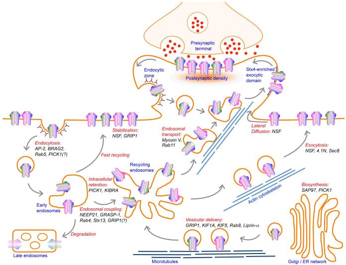
AMPA receptors (AMPARs) mediate the majority of fast excitatory synaptic transmission in the brain. Dynamic changes in neuronal synaptic efficacy, termed synaptic plasticity, are thought to underlie information coding and storage in learning and memory. One major mechanism that regulates synaptic strength involves the tightly regulated trafficking of AMPARs into and out of synapses. The life cycle of AMPARs from their biosynthesis, membrane trafficking, and synaptic targeting to their degradation are controlled by a series of orchestrated interactions with numerous intracellular regulatory proteins. Here we review recent progress made toward the understanding the regulation of AMPAR trafficking, focusing on the roles of several key intracellular AMPAR interacting proteins.


Figures

Figure 1
Figure 1
Structure of AMPAR and its direct interacting proteins. AMPAR is a tetrameric channel assembled from two dimers of different subunits, such as GluA1/GluA2 and GluA2/GluA3. Each individual subunit is composed of a large extracellular ligand-binding domain and a short intracellular carboxyl-tail linked by four transmembrane domains. GluA1 C-terminal domain contains type I PDZ ligand and directly interacts with SAP97, whereas GluA2 C-terminal domain contains type II PDZ ligand and interacts directly with PICK1 and GRIP1. In addition, GluA1 also interacts with protein 4.1N through its juxtamembrane region of the C-terminus, while GluA2 interacts with AP-2, NSF and BRAG-2 through it C-terminus in a non PDZ-dependent manner. Direct binding of AMPARs and these interacting proteins regulates various steps in AMPAR trafficking.
Figure 2
Figure 2
Routes of AMPAR trafficking. AMPARs are assembled in the endoplasmic reticulum and Golgi apparatus in the soma and are delivered into the dendrite via kinesin-dependent vesicular trafficking on microtubule networks prior to their insertion to the plasma membrane. Via lateral diffusion, surface AMPARs are incorporated into synapse and stabilized by postsynaptic density scaffolding proteins. Mature AMPARs undergo constitutive recycling through endosomal trafficking pathway. AMPARs are internalized from the plasma membrane by clathrin-mediated endocytosis and traffic to the early endosome. From early endosome, AMPARs can be delivered back to the plasma membrane either directly (fast recycling) or through recycling endosome, or entering the degradation pathway through late endosome. During LTD, the rate of AMPAR internalization outweighs the rate of AMPAR exocytosis, resulting in reduced number of synaptic AMPARs. Depending on the LTD stimulus, internalized AMPARs can either be retained in intracellular compartment or be degraded in lysosome. Conversely, during LTP, AMPARs are constantly delivered to the plasma membrane to induce early burst and long-term maintenance of synaptic potentiation. Under certain LTP stimuli, recycling endosomes containing AMPARs are directly inserted into dendritic spine exocytic domain, marked by the presence of t-SNARE syntaxin-4. The dendritic spine microdomain also includes a specialized endocytic zone, where AMPARs are rapidly internalized and recycled to provide a large pool of AMPARs during LTP. These highly complex pathways of AMPAR trafficking are tightly regulated by a series of orchestrated interactions with key intracellular regulatory molecules. Disruption of AMPAR binding to its interacting proteins shown in this diagram often leads to aberrant AMPAR trafficking, impaired synaptic plasticity and deficits in learning and memory.

References

    1. Shepherd JD, Huganir RL. The cell biology of synaptic plasticity: AMPA receptor trafficking. Annu Rev Cell Dev Biol. 2007;23:613–643. - PubMed
    1. Henley JM, Barker EA, Glebov OO. Routes, destinations and delays: recent advances in AMPA receptor trafficking. Trends Neurosci. 2011;34:258–268. - PMC - PubMed
    1. Lu W, Shi Y, Jackson AC, Bjorgan K, During MJ, Sprengel R, Seeburg PH, Nicoll RA. Subunit composition of synaptic AMPA receptors revealed by a single-cell genetic approach. Neuron. 2009;62:254–268. * This study provides a molecular quantification of both synaptic and extrasynaptic subunit composition of AMPARs in hippocampal CA1 pyramidal neurons.

    1. Kessels HW, Malinow R. Synaptic AMPA receptor plasticity and behavior. Neuron. 2009;61:340–350. - PMC - PubMed
    1. Correia SS, Bassani S, Brown TC, Lise MF, Backos DS, El-Husseini A, Passafaro M, Esteban JA. Motor protein-dependent transport of AMPA receptors into spines during long-term potentiation. Nat Neurosci. 2008;11:457–466. - PubMed

Publication types

Substances